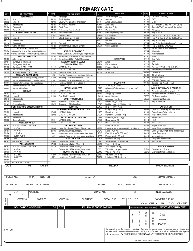
What is the ICD-10 code for K35 80?
ICD-10-CM Code for Unspecified acute appendicitis K35. 80.
What is the ICD-10 code for abnormal appendix?
9: Disease of appendix, unspecified.
What is the icd10 code for appendicitis?
9 Disease of appendix, unspecified.
What is ICD-10 code K37?
ICD-10 code: K37 Unspecified appendicitis | gesund.bund.de.
What is the ICD 10 code for dilated appendix?
K38. 0 is a billable/specific ICD-10-CM code that can be used to indicate a diagnosis for reimbursement purposes. The 2022 edition of ICD-10-CM K38.
What is a dilated appendix?
Mucocele of the appendix is a term used to describe a dilated, mucin-filled appendix. It is most commonly the result of epithelial proliferation, but can be caused by inflammation or obstruction of the appendix.
What is the diagnosis for ICD 10 code r50 9?
9: Fever, unspecified.
What is ICD 10 code R12?
ICD-10 code R12 for Heartburn is a medical classification as listed by WHO under the range - Symptoms, signs and abnormal clinical and laboratory findings, not elsewhere classified .
What is unspecified Acute appendicitis?
Clinical Information. A disorder characterized by acute inflammation to the vermiform appendix caused by a pathogenic agent.
What is a K37?
Appendicitis (pneumococcal) (retrocecal) K37.
What K57 92?
ICD-10 code: K57. 92 Diverticulitis of intestine, part unspecified, without perforation, abscess or bleeding.
How do you code appendectomy in ICD-10?
While 44950 and 44970 stand for open primary appendectomies, 44960 indicates appendectomy for a perforated or ruptured appendix and/or for diffuse peritonitis (ICD-10 code K35. 2).
What is the ICD-10 PCS code for laparoscopic appendectomy?
Excision of Appendix, Percutaneous Endoscopic Approach ICD-10-PCS 0DBJ4ZZ is a specific/billable code that can be used to indicate a procedure.
What is the CPT code for appendicitis?
Two codes differentiate an open appendectomy without rupture (44950) and with rupture (44960). However, only one code applies to laparoscopic appendectomy (44970), and it is used to report a laparoscopic appendectomy for either scenario; with rupture or without rupture (see Table 2, page 43).
What is the ICD-10 for abdominal pain?
ICD-10 code R10. 9 for Unspecified abdominal pain is a medical classification as listed by WHO under the range - Symptoms, signs and abnormal clinical and laboratory findings, not elsewhere classified .
What is the assignment of a diagnosis code?
The assignment of a diagnosis code is based on the provider’s diagnostic statement that the condition exists. The provider’s statement that the patient has a particular condition is sufficient. Code assignment is not based on clinical criteria used by the provider to establish the diagnosis.
What is the convention of ICd 10?
The conventions for the ICD-10-CM are the general rules for use of the classification independent of the guidelines. These conventions are incorporated within the Alphabetic Index and Tabular List of the ICD-10-CM as instructional notes.
What is the ICD-10-CM?
The ICD-10-CM has two types of excludes notes. Each type of note has a different definition for use but they are all similar in that they indicate that codes excluded from each other are independent of each other.
What are the guidelines for coding?
The guidelines are organized into sections. Section I includes the structure and conventions of the classification and general guidelines that apply to the entire classification, and chapter-specific guidelines that correspond to the chapters as they are arranged in the classification. Section II includes guidelines for selection of principal diagnosis for non-outpatient settings. Section III includes guidelines for reporting additional diagnoses in non-outpatient settings. Section IV is for outpatient coding and reporting. It is necessary to review all sections of the guidelines to fully understand all of the rules and instructions needed to code properly.
What is the ICd 10 code for appendix disease?
Disease of appendix, unspecified 1 K38.9 is a billable/specific ICD-10-CM code that can be used to indicate a diagnosis for reimbursement purposes. 2 The 2021 edition of ICD-10-CM K38.9 became effective on October 1, 2020. 3 This is the American ICD-10-CM version of K38.9 - other international versions of ICD-10 K38.9 may differ.
When will the ICD-10-CM K38.9 be released?
The 2022 edition of ICD-10-CM K38.9 became effective on October 1, 2021.
What is the ultrasound of the appendix?
The ultrasound is the preliminary diagnostic tool and may be decisive for the differential diagnosis of appen diceal mu cocele and acute appendicitis. The appendicular diameters of ≥15 mm are the threshold for diagnosing appendiceal mucocele, (sensitivity of 83% specificity of 92%) versus 6-mm outer diameter for the diagnosis of acute appendicitis. [ 3, 8] At the ultrasound, mucocele appears as an elliptical cystic mass with or without acoustic shadowing from dystrophic mural calcification. A mucocele is usually encapsulated on ultrasound, with variable echogenicity in relation to the quantity and fluidity of the mucous contained. The inner wall can appear irregular due to the presence of debris or epithelial hyperplasia. The lumen of giant mucocele can have echogenic layers surrounded by mucin so-called “onion skin sign,” pathognomonic for mucocele. [ 9] Lesser dilated portion of appendix can give “drumstick or pear-shaped” appearance. On ultrasound, mucinous ascites show low-level echoes and poorly defined septation. Ultrasound-guided Fine needle aspiration has not been proposed to avoid dissemination of the mucous leading to pseudomyxoma peritonei.
What is the term for the transformation of the appendix into a mucus-filled sac?
Appendicular mucocele (AM) is the chronic transformation of the appendix into a mucus-filled sac
What causes mucocele in the appendix?
Histopathologically, “mucocele” of the appendix may be caused by a variety of benign and malignant causes of obstruction of the lumen which lead to overproduction of mucus. It is subclassified depending on the depth of invasion of the wall, cellular atypia, extra-appendiceal mucin, and the presence of signet ring cells (associated with poor prognosis). Modern classification is divided into nonneoplastic variants including mucous retention cyst and mucosal hyperplasia and neoplastic variants, including mucosal adenoma (confined to mucosa, mild to moderate cytologic atypia and no atypical mitotic figures), low-grade mucinous neoplasm (low-grade atypia, the loss of muscularis mucosae, and or extra-appendiceal cells, mucocele rupture with extra-appendiceal mucin can lead to this) and mucinous cystadenocarcinoma (high-grade cytologic atypia) [ Table 1 ]. Most benign mucoceles are due to mucinous adenoma which is sessile with dilated appendix with circumferential involvement of mucosa, without extra-appendiceal mucin and are asymptomatic with no recurrence risk after complete excision. Nonneoplastic variant results from chronic, long-standing obstruction of the appendiceal lumen by any nonneoplastic process and are called inflammatory, obstructive, simple mucocele, or retention cyst of the appendix. Mucinous neoplasm of the appendix of low malignant potential is one of the most common causes of “pseudomyxoma peritonei” associated with mucinous peritoneal implants and leads to extensive peritoneal disease without associated lymph node, lung, or liver metastases. [ 4, 5]
What is an appendix mucocele?
Appendicular mucocele (AM) refers to the chronic transformation of the appendix into a mucus-filled sac. It is generally detected in the fifth–seventh decades of life. [ 1] It accounts for 0.3%–0.7% of all appendiceal pathologies and 8% of malignancies of appendix, although has an uncertain histopathological diagnosis, and the terminology is unsettled. [ 2] The clinical presentation is often nonspecific right lower quadrant pain; [ 3] therefore, it is imperative for the clinician to be well aware of differentials. Management with simple appendectomy or right hemicolectomy depends on the preoperative diagnosis of the benign or malignant process. [ 4]
Is appendix mucocele rare?
Mucocele of the appendix is rare and represent s only the tip of the iceberg of underlying benign and malignant pathological processes. Intraoperative diagnosis is also tricky because the inflammation of the appendix often hides the tumor. The preoperative diagnosis is essential to differentiate appendiceal mucocele from acute appendicitis as the treatment varies from open surgical versus laparoscopic surgical approach and for decreasing intraoperative and postoperative morbidity and mortality rate. We present three cases of appendiceal mucocele. The purpose of this paper is to make the physicians aware of the entity, its associations and the effect on management. This review will provide radiologic and pathologic correlation for the preoperative diagnosis of benign and malignant causative processes and differential diagnostic considerations.
Is a mesenteric cyst a developmental cyst?
Mesenteric cyst is reportedly rare and most often detected incidentally. They can be developmental, acquired, neoplastic or infectious or degenerative. In the second case, there was an incidental cystic structure abutting the tip of appendix which raised the possibility of mucinous deposit with appendicular mucocele.
Is mucocele of appendix a rare disease?
Due to its rarity, mucocele of appendix continues to fascinate the surgeon as well as the radiologist and pathologist likewise. It often causes a diagnostic dilemma in the setting of nonspecific symptoms. Although US is often the primary diagnostic modality that is performed, CT is the modality of choice for better characterization. Although a rare disease, surgical management is mandatory because the risk of malignant transformation and prevention of pseudomyxoma peritonei due to rupture of the mucocele itself. Therefore, preoperative diagnosis or suspicion is required for carefully planned resection to excise the tumor.

Popular Posts:
- 1. icd 10 code for unspecified renal insufficiency
- 2. icd 10 code for tobacco use
- 3. icd 10 code for ceruminosis
- 4. icd 10 code for left tibial fibular fracture
- 5. icd 10 code for growth inside lip
- 6. what is the icd 10 code for myelomalacia
- 7. icd 10 cm code for shoved him into the bar table
- 8. icd 10 code for nose congestion
- 9. icd-10 code for foley catheter complications
- 10. icd 10 code for tall stature