What is the ICD 10 code for hemangioma?
Hemangioma of other sites. D18.09 is a billable/specific ICD-10-CM code that can be used to indicate a diagnosis for reimbursement purposes. The 2019 edition of ICD-10-CM D18.09 became effective on October 1, 2018. This is the American ICD-10-CM version of D18.09 - other international versions of ICD-10 D18.09 may differ.
What is the ICD 10 code for hepatomegaly?
2021 ICD-10-CM Diagnosis Code R16.0 Hepatomegaly, not elsewhere classified 2016 2017 2018 2019 2020 2021 Billable/Specific Code R16.0 is a billable/specific ICD-10-CM code that can be used to indicate a diagnosis for reimbursement purposes.
What is the ICD 10 code for benign neoplasm of liver?
Benign neoplasm of liver 2016 2017 2018 2019 2020 2021 Billable/Specific Code D13.4 is a billable/specific ICD-10-CM code that can be used to indicate a diagnosis for reimbursement purposes. The 2021 edition of ICD-10-CM D13.4 became effective on October 1, 2020.
What is the ICD 10 code for hepatic coma?
ICD-10-CM Diagnosis Code D18.01. Hemangioma of skin and subcutaneous tissue. 2016 2017 2018 2019 2020 2021 Billable/Specific Code. ICD-10-CM Diagnosis Code B15.0 [convert to ICD-9-CM] Hepatitis A with hepatic coma. Viral hepatitis a with hepatic coma. ICD-10-CM Diagnosis Code B15.0. Hepatitis A with hepatic coma.
What is the ICD-10 code for hepatic hemangioma?
ICD-10-CM Code for Hemangioma D18. 0.
What is the ICD-10 code for hemangioma?
ICD-10 code D18. 01 for Hemangioma of skin and subcutaneous tissue is a medical classification as listed by WHO under the range - Neoplasms .
What is the ICD-10 code for right hepatic lobe lesion?
K76. 89 is a billable/specific ICD-10-CM code that can be used to indicate a diagnosis for reimbursement purposes. The 2022 edition of ICD-10-CM K76. 89 became effective on October 1, 2021.
What is a hepatic hemangioma of the liver?
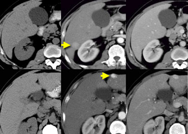
A hepatic hemangioma is a liver mass made of widened (dilated) blood vessels. It is not cancerous. This upper abdominal CT scan shows a blood vessel tumor (hemangioma) in the liver. The digestive system organs in the abdominal cavity include the liver, gallbladder, stomach, small intestine and large intestine.
What is the ICD-10 code for liver mass?
There are four different ICD-10 diagnosis codes for the four conditions listed above. For example, a liver lesion is coded as K76. 9; a liver mass is coded as R16. 0, a liver tumor is coded as D49.
What hemangioma mean?
A hemangioma (hee man jee OH mah) is a common vascular birthmark, made of extra blood vessels in the skin. It is a benign (non-cancerous) growth. The exact cause is not known. Hemangiomas are typically not inherited, but others in the family may also have had them.
What are hepatic lesions?
Liver lesions are cell abnormalities within the liver. They are most frequently benign, but some can be cancerous. Liver lesions can be caused due to a variety of reasons. Most of the lesions don't cause any symptoms until they develop into larger masses.
What is diagnosis code R16?
Hepatomegaly and splenomegaly, not elsewhereICD-10 code R16 for Hepatomegaly and splenomegaly, not elsewhere classified is a medical classification as listed by WHO under the range - Symptoms, signs and abnormal clinical and laboratory findings, not elsewhere classified .
What is hepatic mass?
Hepatic tumors are a diverse group of masses that include malignant and benign subtypes. Their presentation can vary from localizing signs/symptoms, such as jaundice and right upper quadrant pain, to vague signs/symptoms, such as fatigue, weight loss, and anorexia.
What causes hepatic hemangiomas?
What causes a liver hemangioma? Doctors are still not sure what causes liver hemangiomas. In some cases, liver hemangiomas may be present from birth, but they can also develop at any point during a person's life. They are more common in people aged 30–50 years, and more likely to occur in women than in men.
Is a liver hemangioma a tumor?
Hemangiomas are bundles of blood vessels that form benign, or noncancerous, tumors in the liver. Many people who have liver hemangiomas don't know they have them.
Is a hemangioma a mass?
They can be small or large in size and may be flat to the skin, raised, or protrude out as a nodule. Some appear as a spongy mass that covers an entire extremity (called "diffuse hemangioma" or "angiomatosis"). Cavernous hemangioma.
Is hemangioma in liver serious?
The hemangioma, or tumor, is a tangle of blood vessels. It's the most common noncancerous growth in the liver. It's rarely serious and doesn't turn into liver cancer even when you don't treat it.
Should I be worried about liver hemangioma?
If your liver hemangioma is small and doesn't cause any signs or symptoms, you won't need treatment. In most cases a liver hemangioma will never grow and will never cause problems. Your doctor may schedule follow-up exams to check your liver hemangioma periodically for growth if the hemangioma is large.
How do you treat a hemangioma in the liver?
Hepatic hemangiomas have been treated with a wide array of therapies. Traditionally, surgical resection and surgical enucleation are the treatments of choice. Minimally invasive therapies for hepatic hemangioma include arterial embolization, radiofrequency ablation, and hepatic irradiation.
Is hemangioma serious?
If left untreated, symptomatic hemangiomas can cause serious neurological effects. At UPMC, we treat hemangiomas with surgical removal (resection) of the tumor or the affected vertebra, and radiation therapy to treat pain.
The ICD code D180 is used to code Capillary hemangioma
A capillary hemangioma (also known as an Infantile hemangioma, Strawberry hemangioma,:593 and Strawberry nevus) is the most common variant of hemangioma which appears as a raised, red, lumpy area of flesh anywhere on the body, though 83% occur on the head or neck area.
Coding Notes for D18.0 Info for medical coders on how to properly use this ICD-10 code
Inclusion Terms are a list of concepts for which a specific code is used. The list of Inclusion Terms is useful for determining the correct code in some cases, but the list is not necessarily exhaustive.
ICD-10-CM Alphabetical Index References for 'D18.0 - Hemangioma'
The ICD-10-CM Alphabetical Index links the below-listed medical terms to the ICD code D18.0. Click on any term below to browse the alphabetical index.
What is the code for a primary malignant neoplasm?
A primary malignant neoplasm that overlaps two or more contiguous (next to each other) sites should be classified to the subcategory/code .8 ('overlapping lesion'), unless the combination is specifically indexed elsewhere.
What chapter is neoplasms classified in?
All neoplasms are classified in this chapter, whether they are functionally active or not. An additional code from Chapter 4 may be used, to identify functional activity associated with any neoplasm. Morphology [Histology] Chapter 2 classifies neoplasms primarily by site (topography), with broad groupings for behavior, malignant, in situ, benign, ...
Popular Posts:
- 1. icd-10 code for acute pain
- 2. icd 10 code for type 1 neurofibromatosis
- 3. icd 10 code for umbilical vein varix in pregnancy
- 4. icd 10 code for left shoulder decreased range of motion
- 5. icd 10 code for degenerative osteo arthritis hip
- 6. icd-10 code for phyical abuse of child
- 7. icd 10 code for subcutaneous abscess abdominal wall
- 8. icd 10 code for immune compromised
- 9. icd 10 code for conn's syndrome
- 10. icd 10 code for iron deficiency anemia due to menorrhagia