How to code medical diagnosis?
- Point out the tests that were already performed to show the reason for the problem.
- Explain how these evaluations confirmed your diagnosis and show conclusive evidence.
- Use factual information, such as test result quotes, to back up your identification of the patient's issue.
What are DSM diagnosis codes?
Mental retardation
- 317 Mild mental retardation
- 318.0 Moderate mental retardation
- 318.1 Severe mental retardation
- 318.2 Profound mental retardation
- 319 Mental retardation; severity unspecified
What do these diagnosis codes mean?
The CPT code describes what was done to the patient during the consultation, including diagnostic, laboratory, radiology, and surgical procedures while the ICD code identifies a diagnosis and describes a disease or medical condition. … CPT codes are more complex than ICD codes. What is a procedure code and why is it used?
What does diagnosis code 78079 mean?
ICD-9-CM 780.79 is a billable medical code that can be used to indicate a diagnosis on a reimbursement claim, however, 780.79 should only be used for claims with a date of service on or before September 30, 2015. For claims with a date of service on or after October 1, 2015, use an equivalent ICD-10-CM code (or codes).

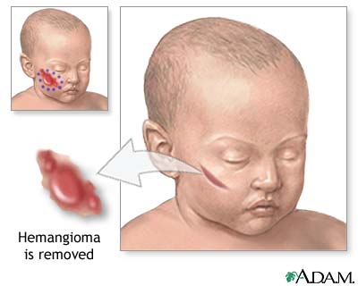
What is a hemangioma tumor?
A hemangioma (he-man-jee-O-muh) is a bright red birthmark that shows up at birth or in the first or second week of life. It looks like a rubbery bump and is made up of extra blood vessels in the skin. A hemangioma can occur anywhere on the body, but most commonly appears on the face, scalp, chest or back.
What is the ICD-10 code for hepatic hemangioma?
D18. 09 is a billable/specific ICD-10-CM code that can be used to indicate a diagnosis for reimbursement purposes. The 2022 edition of ICD-10-CM D18. 09 became effective on October 1, 2021.
What is the ICD-10 code for hemangioma of scalp?
D18. 01 - Hemangioma of skin and subcutaneous tissue | ICD-10-CM.
What is the ICD-10-CM code for a cavernous hemangioma?
02.
What is hemangioma of intra abdominal structures?
They are benign tumours that arise from embryonic remnants of unipotent angioblastic cells [1]. Although hemangiomas may occur anywhere within the abdomen, including the solid organs, hollow viscera, ligaments, and abdominal wall, the liver is the most common site.
What is the ICD-10-CM code for a cavernous hemangioma in intracranial structures?
02.
What is hemangioma of skin and subcutaneous tissue?
Hemangiomas of the skin can form in the top layer of skin or in the fatty layer underneath, which is called the subcutaneous layer. At first, a hemangioma may appear to be a red birthmark on the skin. Slowly, it will start to protrude upward from the skin. However, hemangiomas are not usually present at birth.
Is Angioma the same as hemangioma?
Angioma or haemangioma (American spelling 'hemangioma') describes a benign vascular skin lesion. An angioma is due to proliferating endothelial cells; these are the cells that line the inside of a blood vessel.
What causes a hemangioma?
Key points about hemangiomas and vascular malformations Hemangiomas and vascular malformations are noncancerous growths. They show up at birth or soon after birth. They're also called birthmarks. The cause of these growths often isn't known.
What is a cavernous angioma?
A cavernoma is a cluster of abnormal blood vessels, usually found in the brain and spinal cord. They're sometimes known as cavernous angiomas, cavernous hemangiomas, or cerebral cavernous malformation (CCM). A typical cavernoma looks like a raspberry.
What is the ICD 10 code for Lipoma?
D17.22 for Benign lipomatous neoplasm of skin and subcutaneous tissue of limb is a medical classification as listed by WHO under the range - Neoplasms .
What is the ICD 10 code for Cavernoma?
Q28. 3 - Other malformations of cerebral vessels | ICD-10-CM.
What is a benign skin lesion?
The majority of cases are congenital. A benign skin lesion consisting of dense, usually elevated masses of dilated blood vessels. A benign tumor of the blood vessels that appears on skin. A benign vascular neoplasm characterized by the formation of capillary-sized or cavernous vascular channels.
What chapter is functional activity?
Functional activity. All neoplasms are classified in this chapter, whether they are functionally active or not. An additional code from Chapter 4 may be used, to identify functional activity associated with any neoplasm. Morphology [Histology]
Is morphology included in the category and codes?
In a few cases, such as for malignant melanoma and certain neuroendocrine tumors, the morphology (histologic type) is included in the category and codes. Primary malignant neoplasms overlapping site boundaries.
What is a benign vascular neoplasm characterized by the formation of capillary-sized or
A benign vascular neoplasm characterized by the formation of capillary-sized or cavernous vascular channels. A hemangioma characterized by the presence of cavernous vascular spaces. A vascular anomaly due to proliferation of blood vessels that forms a tumor-like mass.
What is the code for a primary malignant neoplasm?
A primary malignant neoplasm that overlaps two or more contiguous (next to each other) sites should be classified to the subcategory/code .8 ('overlapping lesion'), unless the combination is specifically indexed elsewhere.
What is the table of neoplasms used for?
The Table of Neoplasms should be used to identify the correct topography code. In a few cases, such as for malignant melanoma and certain neuroendocrine tumors, the morphology (histologic type) is included in the category and codes. Primary malignant neoplasms overlapping site boundaries.
What chapter is functional activity?
Functional activity. All neoplasms are classified in this chapter, whether they are functionally active or not. An additional code from Chapter 4 may be used, to identify functional activity associated with any neoplasm. Morphology [Histology]

Popular Posts:
- 1. icd-10-cd code(s) for congenital cystic kidney disease
- 2. icd 10 cm code for short term memory loss
- 3. icd 10 code for high risk pregnancy
- 4. icd 10 code for trigger finger right ring finger
- 5. what is the icd 10 code for back riyria
- 6. icd 10 code for c22.9
- 7. icd 9 code for 278.00
- 8. icd 10 code for abnormal bleeding for pregnant woman
- 9. icd-10 code for mammogram screening
- 10. icd 10 code for left ischial tuberosity ulcer