59 Anterior Corneal Dystrophies. Epithelial basement membrane dystrophy (EBMD) is a degenerative condition of the anterior layer of the cornea.Aug 6, 2016
What is the ICD 10 code for basement membrane dystrophy?
ICD-10 Diagnosis Code: Epithelial basement membrane dystrophy (EBMD) is a degenerative condition of the anterior layer of the cornea. DEFINITION. Epithelial basement membrane dystrophy (EBMD) is characterized by abnormal quantities of basement membrane and cytoplasmic debris that are misdirected into the corneal epithelium.
What is the classification of epithelial basement membrane dystrophy?
CLASSIFICATION. According to the International Committee for Classification of Corneal Dystrophies (IC3D), epithelial basement membrane dystrophy is a Categoy 1 Corneal Dystrophy. In this type of dystrophy, the gene has been identified and mapped and specific mutations are known.
What is the ICD-10 code for Mat-dot-frequency (MDF) dystrophy?
Question: What ICD-10 code do you recommend for Mat-Dot-Frequency (MDF) dystrophy? I'm told Anterior basement membrane dystrophy (ABMD) and MDF are very similar. Answer: The ICD-10-CM for Ophthalmology: The Complete Reference maps 371.52 Map- dot fingerprint corneal dystrophy to H18.59 Other hereditary corneal dystrophies.
What is the ICD 10 code for corneal dystrophy?
ICD-10 Diagnosis Code: H18.59–Other hereditary corneal dystrophy. Title Anterior Corneal Dystrophies Category Corneal Opacity And Other Disorders Of Cornea. Description Epithelial basement membrane dystrophy (EBMD) is a degenerative condition of the anterior layer of the cornea.
What is anterior basement membrane dystrophy?
Anterior Basement Membrane Corneal Dystrophy is the official name for Map Dot Fingerprint Corneal Dystrophy. In this condition, the basement membrane under the corneal epithelium does not function properly. The basement membrane functions as a sticky anchor over which the epithelium grows.
What is epithelial basement membrane dystrophy?
Epithelial basement membrane dystrophy (EBMD) is a disease that affects the anterior cornea, causing characteristic slit lamp findings which may result in decreased vision and/or recurrent corneal erosions.
What is the ICD 10 code for epithelial defect?
Epithelial (juvenile) corneal dystrophy The 2022 edition of ICD-10-CM H18. 52 became effective on October 1, 2021.
Is epithelial basement membrane dystrophy rare?
Epithelial basement membrane dystrophy is a common form of corneal dystrophy and is also known as map-dot-fingerprint dystrophy and Cogan microcystic dystrophy. This extremely rare form of corneal dystrophy affects the epithelial layer of the cornea.
Where is basement membrane?
The basement membrane (BM) is a special type of extracellular matrix that lines the basal side of epithelial and endothelial tissues.
Are Abmd and EBMD the same?
Epithelial Basement Membrane Dystrophy (EBMD), is the most common of the corneal dystrophies. Ii is also known as Map-Dot-Fingerprint Dystrophy and Anterior Basement Membrane Dystrophy (ABMD), . Since it was first described by Cogan et.al. in 1964, it is also known as Cogan's Microcystic Corneal Dystrophy.
What is the ICD-10 code for epithelial basement membrane dystrophy?
59 Anterior Corneal Dystrophies. Epithelial basement membrane dystrophy (EBMD) is a degenerative condition of the anterior layer of the cornea.
What is an epithelial defect?
Corneal epithelial defects are focal areas of epithelial (outermost corneal layer) loss; they can be due to mechanical trauma, corneal dryness, neurotrophic disease, post surgical changes, infection, or any other of a variety of etiologies.
What is the ICD-10 code for epiretinal membrane?
For documentation of epiretinal membrane, follow Index lead term Disease/retina/specified NEC to assign H35. 8 Other specified retinal disorders.
Is anterior basement membrane dystrophy hereditary?
Anterior Basement Membrane Dystrophy (ABMD) is an inherited disorder of the cornea that may present with a variety of symptoms, including recurrent corneal erosions and/or blurred vision.
What is the difference between degeneration and dystrophy?
Degenerations are usually unilateral, asymmetric and often peripheral. Changes caused by inflammation, maturity or systemic disease result in deposition, thinning or vascularization of the corneal tissue. Dystrophies are rare conditions and may not present in a primary setting.
Which is the most common corneal dystrophy?
The most common is Fuchs' corneal dystrophy, which usually starts when you're in your 40s or 50s. It may take several more years, even decades before you notice vision problems. With Fuchs', the cells that pump excess moisture out of your cornea to keep it clear start to die.
What is EBMD in medical terms?
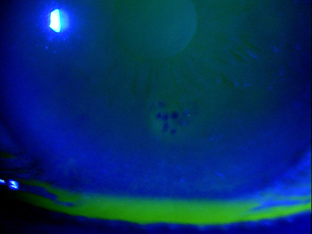
Epithelial basement membrane dystrophy (EBMD) is characterized by abnormal quantities of basement membrane and cytoplasmic debris that are misdirected into the corneal epithelium. Clinically, the abnormal deposits in EBMD appear as dot-like opacities, map-like patterns, or whorled fingerprint-like lines in the corneal epithelium. In many patients, the epithelial lesions change in appearance, location and number over time.
What is the anterior layer of the cornea?
The anterior layer of the cornea is composed of the corneal epithelium and its underlying basement membrane. The basal cells of the corneal epithelium produce and adhere to the basement membrane via hemidesmosomes and basement membrane complexes.
Can EBMD cause symptoms?
Patient symptoms vary depending on the severity of the disease and mild conditions may produce no symptoms. Patients with EBMD may present with any of the following abnormal clinical signs and symptoms:
Popular Posts:
- 1. 2019 icd 10 code for matched defect within the right middle cerebral artery territory
- 2. icd-10 code for dislocation of right hip prosthesis
- 3. icd 10 code for trauma to the foot
- 4. 2020 icd 10 code for buttock pain
- 5. icd 10 code for acute epidural hematoma
- 6. icd 10 cm code for postoperative peritoneal adhesions
- 7. icd-10 code for blocked tear duct in infant
- 8. icd 10 code for nash cirrhosis
- 9. icd-10 code for hashimoto's disease
- 10. icd 9 code for pulmonary emboli