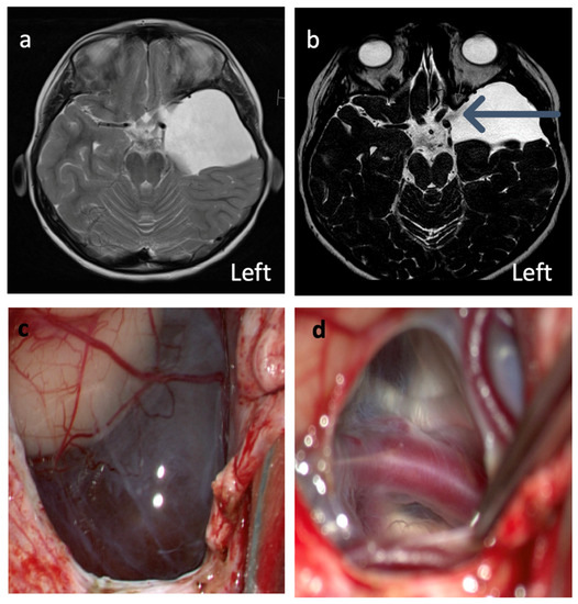
What is the treatment for arachnoid cyst?
What You Need to Know
- Arachnoid cysts occur in one of the three layers of tissue that surround the brain and spinal cord.
- Most arachnoid cysts are stable and do not require treatment.
- They are four times more common in boys than in girls.
- Arachnoid cysts are diagnosed with a CT or MRI scan.
- Treatment, if necessary, involves draining the fluid through surgery or shunting.
What does arachnoid cysts mean?
Arachnoid cysts are cerebrospinal fluid-filled sacs that are located between the brain or spinal cord and the arachnoid membrane, one of the three membranes that cover the brain and spinal cord. Primary arachnoid cysts are present at birth and are the result of developmental abnormalities in the brain and spinal cord that arise during the early weeks of gestation.
Do arachnoid cyst is the cause for the strokes?
Untreated arachnoid cysts may cause permanent, severe neurological damage due to expansion of the cyst, hydrocephalus, or hemorrhage. With treatment, most individuals with arachnoid cysts have a good prognosis. Symptoms of an arachnoid cyst depend on the cyst’s size and location.
Do arachnoid cyst cause nose bleeds?
Symptoms often resolve or improve with treatment but if left untreated, arachnoid cysts may cause permanent severe neurological damage due to the progressive expansion of the cyst (s) or bleeding into the cyst. Arachnoid cysts may also result in a subdural hematoma, most commonly after a head trauma.

What is the ICD-10 code for arachnoid cyst?
Other disorders of meninges, not elsewhere classified The 2022 edition of ICD-10-CM G96. 19 became effective on October 1, 2021. This is the American ICD-10-CM version of G96. 19 - other international versions of ICD-10 G96.
What is an arachnoid cyst?
Arachnoid cysts are cerebrospinal fluid-filled sacs that are located between the brain or spinal cord and the arachnoid membrane, one of the three membranes that cover the brain and spinal cord.
Is an arachnoid cyst a lesion?
What are arachnoid cysts? Arachnoid cysts are fluid-filled sacs that grow on the brain and spine. They are not tumors, and they are not cancerous. On rare occasions, if they grow too big or press on other structures in the body, they can cause brain damage or movement problems.
What is the ICD-10 code for colloid cyst?
In the ICD-10-CM code book, locate the term “cyst” in the index, followed by the term “brain” and look down to the terms of “third ventricle (colloid), congenital” to obtain the code Q04. 6.
Is an arachnoid cyst a brain tumor?
The cysts are fluid-filled sacs, not tumors. The likely cause is a split of the arachnoid membrane, one of the three layers of tissue that surround and protect the brain and spinal cord.
Why is it called an arachnoid cyst?
An arachnoid cyst is most likely to develop in your head, but it can also develop around your spinal cord. It's called an arachnoid cyst because it occurs in the space between your brain, or spinal column, and your arachnoid membrane. This is one of three membrane layers that surround your brain and spine.
Is a brain cyst the same as a tumor?
Brain cysts located in the brain are not truly “brain tumors” because they do not arise from the brain tissue itself. Although they tend to be (benign noncancerous), they are sometimes found in parts of the brain that control vital functions.
How common is an arachnoid cyst?
In the United States, about 3 children in every 100 have an arachnoid cyst. Most of these cysts never cause any problems or symptoms or need any treatment. Doctors often find arachnoid cysts when they examine a child for another reason, such as after a head injury.
What is a cyst in the brain?
A brain cyst or cystic brain lesion is a fluid-filled sac in the brain. They can be noncancer (benign) or cancer (malignant). Benign means that the growth doesn't spread to other parts of the body. A cyst may contain blood, pus, or other material. In the brain, cysts sometimes contain cerebrospinal fluid (CSF).
What is the ICD-10 code for brain tumor?
ICD-10-CM Code for Malignant neoplasm of brain, unspecified C71. 9.
What is fenestration of arachnoid cyst?
Endoscopic cyst fenestration is a minimally invasive technique in which a burr hole is made and an endoscope is guided through the opening. Upon reaching the cyst, the neurosurgeon fenestrates the walls of the cyst, draining the fluid. It is the procedure of choice for most instances of arachnoid cysts.
What is a colloid cyst?
A colloid cyst is a slow-growing tumor typically found near the center of the brain. If large enough, a colloid cyst obstructs cerebrospinal fluid (CSF) movement, resulting in a build up of CSF in the ventricles of the brain (hydrocephalus) and elevated brain pressure.
Popular Posts:
- 1. icd 10 cm code for stuck by vehicle accident
- 2. icd 10 code for infusing an antibiotic
- 3. icd 10 code for bradycardia palpitations
- 4. icd 10 code for dilated pore
- 5. icd 10 code for iliac artery stent stenosis
- 6. icd 10 code for egd
- 7. icd-10-cm code for htn with heart failure
- 8. icd 10 cm code for gonorrhea test
- 9. icd 10 code for no prenatal care
- 10. icd 10 code for left trimalleolar fracture