Atrophic flaccid tympanic membrane, unspecified ear
H73. 819 is a billable/specific ICD-10-CM code that can be used to indicate a diagnosis for reimbursement purposes. The 2022 edition of ICD-10-CM H73. 819 became effective on October 1, 2021.What is the ICD 10 code for atelectasis?
Atelectasis. J98.11 is a billable/specific ICD-10-CM code that can be used to indicate a diagnosis for reimbursement purposes. The 2020 edition of ICD-10-CM J98.11 became effective on October 1, 2019. This is the American ICD-10-CM version of J98.11 - other international versions of ICD-10 J98.11 may differ.
What is the ICD 10 code for ear infection?
Other specified disorders of ear, unspecified ear. H93.8X9 is a billable/specific ICD-10-CM code that can be used to indicate a diagnosis for reimbursement purposes. The 2020 edition of ICD-10-CM H93.8X9 became effective on October 1, 2019.
What is the ICD 10 code for eardrum prolapse?
ICD-10-CM Diagnosis Code H66.01 ICD-10-CM Diagnosis Code S09.2 "Includes" further defines, or give examples of, the content of the code or category. A temporary or persistent opening in the eardrum (tympanic membrane). Clinical signs depend on the size, location, and associated pathological condition.
What is the ICD 10 code for blocked ears?
Sensation of blocked ears. ICD-10-CM H93.8X9 is grouped within Diagnostic Related Group (s) (MS-DRG v38.0): 154 Other ear, nose, mouth and throat diagnoses with mcc. 155 Other ear, nose, mouth and throat diagnoses with cc.
What is the ICD-10 diagnosis code for atelectasis?
ICD-10 code J98. 11 for Atelectasis is a medical classification as listed by WHO under the range - Diseases of the respiratory system .
What is retracted tympanic membrane?
A tympanic membrane retraction, or retracted eardrum, is a condition where the tympanic membrane, or eardrum, gets pulled toward the middle of your ear. The tympanic membrane is a thin layer of tissue found between your inner and outer ear.
Where is your eardrum?
The eardrum divides the outer ear from the middle ear. The eardrum sits between the end of the external ear canal and the auditory ossicles, which are three tiny bones in the middle ear, called the malleus, incus, and stapes.
What is unspecified perforation of tympanic membrane?
What is an eardrum (tympanic membrane) perforation? Tympanic membrane perforation, also known as a perforated eardrum, is a hole in the thin membrane that separates the ear canal from the middle ear.
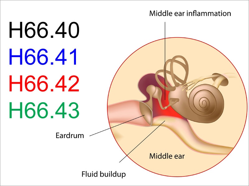
What is middle ear atelectasis?
Atelectasis of the middle ear is a condition of retraction and atrophy of the tympanic membrane in chronic otitis media with effu- sion. The tympanic membrane, in the pres- ence of middle ear effusion, shows a variety of changes.
What is a collapsed ear canal?
Background: Collapsed ear canals typically occur when an outside force, such as a headset for audiometric testing, is present. However, when a collapsed ear canal occurs without external pressure, this creates a challenge not only for performing audiometric testing but also for coupling a hearing aid to the ear canal.
What is the eardrum called?
tympanic membraneThe sound waves then travel toward a flexible, oval membrane at the end of the ear canal called the eardrum, or tympanic membrane. Sound waves cause the eardrum to vibrate.
Is eardrum part of middle ear?
Tympanic membrane (eardrum). The tympanic membrane divides the external ear from the middle ear. Middle ear (tympanic cavity), consisting of: Ossicles.
What is the ear drum?
The tympanic membrane (eardrum) separates the outer ear from the middle ear. The membrane vibrates when sound waves strike it, beginning the process that converts the sound wave into a nerve impulse that travels to the brain. The ear consists of external, middle, and inner structures.
When do you refer to tympanic membrane perforation?
Symptoms include sudden ear pain, or sudden decrease in ear pain, discharge (which may be bloody) or hearing loss. The vast majority of ruptured eardrums will heal without treatment. A simple perforation of the ear drum as part of acute otitis media does NOT need referral unless it persists > 6 weeks.
How do you classify tympanic membrane perforation?
The classification was according to the size of perforation: small perforation, <1 quadrants or maximum diameter <3. mm; middle perforation, >1 quadrants and <2 quadrants or maximum diameter between 3 and 5 mm; and large perforation, >2 quadrants or maximum diameter >5 mm.
What is the most common type of tympanic membrane perforation?
BACKGROUND Tympanic membrane perforations are common and can be categorised into either acute or chronic. Acute perforations are usually traumatic or inflammatory in origin and heal spontaneously. Chronic perforations may be associated with underlying progressive disease.
Can you touch your eardrum?
Puncturing or tearing your eardrum It's possible, but very unlikely, that you'll poke a hole in your eardrum, also known as the tympanic membrane, while cleaning your ears with Q-tips. Your ears have a lot of nerve endings that send powerful feedback to your brain telling you that what you're doing is painful.
How far down is your eardrum?
The adult human ear canal extends from the pinna to the eardrum and is about 2.5 centimetres (1 in) in length and 0.7 centimetres (0.3 in) in diameter....Ear canalTA26867FMA61734Anatomical terminology11 more rows
Can you see the eardrum by just looking in the ear?
The eardrum is pearly white or light gray, and you can see through it. You can see the tiny bones of the middle ear pushing on the eardrum. You see a cone of light, known as the "light reflex," reflecting off the surface of the eardrum.
How do you massage ear wax out?
To do this, just gently massage the outside of the ear using circular movements. That way, the impaction will soften, which can help the earwax drain more easily. Once you've finished making these circular movements, pull your ear slightly backwards, from the lobe to the top of the auricle.
What is the ICD code for atelectasis?
J98.11 is a billable ICD code used to specify a diagnosis of atelectasis. A 'billable code' is detailed enough to be used to specify a medical diagnosis.
What is the ICd 9 code for pulmonary consolidation?
It is a condition where the alveoli are deflated down to little or no volume, as distinct from pulmonary consolidation, in which they are filled with liquid. Specialty: Pulmonology. MeSH Code: D001261. ICD 9 Code: 518.0.

Popular Posts:
- 1. icd=1- code for hemiplegia migraine
- 2. icd 10 code for paralysis of left lower extremity
- 3. icd 10 code for b 12 dif
- 4. icd 10 cm code for fell off her board
- 5. icd 10 code for systolic dysfunction
- 6. icd 10 code for contusion of upper arm
- 7. icd 10 code for chronic t11 compression fracture
- 8. icd-10-pcs code for beam radiation for prostate cancer
- 9. icd 10 cm code for hx of sepsis
- 10. 2016 icd 10 code for calculi