I78. 1 is a billable/specific ICD-10-CM code that can be used to indicate a diagnosis for reimbursement purposes.
What is the ICD-10-CM code for nevus?
2016 2017 2018 2019 2020 2021 2022 Billable/Specific Code POA Exempt. ICD-10-CM Diagnosis Code M84.750G [convert to ICD-9-CM] Atypical femoral fracture, unspecified, subsequent encounter for fracture with delayed healing. Atypical femoral fracture, unspecified, 7thG. ICD-10-CM Diagnosis Code M84.750G.
What is the ICD 10 code for melanocytic nevi?
May 10, 2019 · What is the ICD 10 code for atypical nevus? I78. 1 is a billable/specific ICD-10-CM code that can be used to indicate a diagnosis for reimbursement purposes. Is a melanocytic nevus cancerous? Melanocytic nevi are benign neoplasms or hamartomas composed of melanocytes, the pigment-producing cells that constitutively colonize the epidermis.
What is an atypical nevus?
Apr 01, 2022 · What is the ICD 10 code for atypical mole? Nevus, non-neoplastic. I78. 1 is a billable/specific ICD-10-CM code that can be used to indicate a diagnosis for reimbursement purposes. The 2022 edition of ICD-10-CM I78. What is nevus non neoplastic?
What is the ICD 10 code for non-neoplastic neoplasm?
Atypical chronic myeloid leukemia, BCR/ABL-neg, in remission; Atypical chronic myeloid leukemia, bcr/abl negative in remission; Atypical chronic myeloid leukemia, bcr/abl negative, i ICD-10-CM Diagnosis Code C92.21
What is the ICD-10 code for atypical mole?
ICD-10 | Melanocytic nevi, unspecified (D22. 9)
What is ICD-10 code nevus?
1.
What is melanocytic nevi of scalp and neck?
Melanocytic nevi are benign neoplasms or hamartomas composed of melanocytes, the pigment-producing cells that constitutively colonize the epidermis.Nov 1, 2019
What is diagnosis code D22 5?
Melanocytic nevi of trunk5: Melanocytic nevi of trunk.
What is nevus non neoplastic?
Definition. A abnormal, congenital formation or mark on the skin or neighboring mucosa that does not show neoplastic growth. [ from NCI]
Is a compound nevus benign or malignant?
Compound naevi are benign lesions. They do not cause complications and they have an excellent prognosis.Mar 25, 2022
What is an atypical nevus?
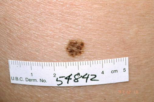
INTRODUCTION. Atypical nevi, also known as dysplastic nevi, are benign acquired melanocytic neoplasms. Atypical nevi share some of the clinical features of melanoma, such as asymmetry, irregular borders, multiple colors, and diameter >5 mm (picture 1A). They occur sporadically or in a familial setting.Sep 16, 2020
What is atypical lentiginous nevus?
Atypical lentiginous nevus (of the elderly) is a peculiar form of dysplastic nevus. Clinically, this condition can resemble malignant melanoma and histologically, it has a lentiginous pattern with variable degrees of atypia and an absence of dermal nests.
What is dermal nevus?
A dermal nevus is a non-cancerous type of growth made up of specialized cells called melanocytes. Dermal nevi (more than one nevus are called nevi) are usually seen in individuals of lighter skin complexion and can be found anywhere on the body.
What is the ICD-10 code for dysplastic nevus?
D22. 9 is a billable/specific ICD-10-CM code that can be used to indicate a diagnosis for reimbursement purposes.
What is ICD-10 code lentigo simplex?
4: Other melanin hyperpigmentation.
What is the ICD-10 code for eczema?
Dermatitis and eczema L20-L30.
What is the color of a nevus?
A dysplastic nevus is often larger with borders that are not easy to see. Its color is usually uneven and can range from pink to dark brown. Parts of the mole may be raised above the skin surface. A dysplastic nevus may develop into malignant melanoma (a type of skin cancer).
What is a Melanocytic Nevi?
A benign (not cancer) growth on the skin that is formed by a cluster of melanocytes (cells that make a substance called melanin, which gives color to skin and eyes). A mole is usually dark and may be raised from the skin.
How many moles are there on the skin?
Moles are very common. Most people have between 10 and 40 moles. A person may develop new moles from time to time, usually until about age 40.moles are usually pink, tan or brown.
What is a mole on the skin?
A mole is usually dark and may be raised from the skin. A circumscribed stable malformation of the skin and occasionally of the oral mucosa, which is not due to external causes and therefore presumed to be of hereditary origin. A neoplasm composed of melanocytes that usually appears as a dark spot on the skin.
What is the code for a primary malignant neoplasm?
A primary malignant neoplasm that overlaps two or more contiguous (next to each other) sites should be classified to the subcategory/code .8 ('overlapping lesion'), unless the combination is specifically indexed elsewhere.
What chapter is neoplasms classified in?
All neoplasms are classified in this chapter, whether they are functionally active or not. An additional code from Chapter 4 may be used, to identify functional activity associated with any neoplasm. Morphology [Histology] Chapter 2 classifies neoplasms primarily by site (topography), with broad groupings for behavior, malignant, in situ, benign, ...
What is a benign growth on the skin?
A benign growth on the skin (usually tan, brown, or flesh-colored) that contain s a cluster of melanocytes and surrounding supportive tissue. A neoplasm composed of melanocytes that usually appears as a dark spot on the skin. A nevus characterised by the presence of excessive pigment. A nevus containing melanin.
What is a mole on the skin?
A mole is a cluster of melanocytes and surrounding supportive tissue that usually appears as a tan, brown, or flesh-colored spot on the skin. The plural of nevus is nevi (nee-vye).
What is a nevus pigmented?
NEVUS PIGMENTED-. a nevus containing melanin. the term is usually restricted to nevocytic nevi round or oval collections of melanin containing nevus cells occurring at the dermoepidermal junction of the skin or in the dermis proper or moles but may be applied to other pigmented nevi.
What are vascular birthmarks?
Vascular birthmarks are made up of blood vessels that haven't formed correctly. They are usually red. Two types of vascular birthmarks are hemangiomas and port-wine stains. Pigmented birthmarks are made of a cluster of pigment cells which cause color in skin.
What are birthmarks on the skin called?
Birthmarks. Also called: Cafe au lait spot, Hemangioma, Mongolian spot, Nevus, Strawberry mark. Birthmarks are abnormalities of the skin that are present when a baby is born. There are two types of birthmarks.
What is the GEM crosswalk?
The General Equivalency Mapping (GEM) crosswalk indicates an approximate mapping between the ICD-10 code D22.9 its ICD-9 equivalent. The approximate mapping means there is not an exact match between the ICD-10 code and the ICD-9 code and the mapped code is not a precise representation of the original code.
How many moles are there in the human body?
They happen when pigment cells in the skin, called melanocytes, grow in clusters. Moles are very common. Most people have between 10 and 40 moles. A person may develop new moles from time to time, usually until about age 40. In older people, they tend to fade away. Moles are usually pink, tan or brown.
Can a mole be a birthmark?
Moles can be birthmarks. No one knows what causes many types of birthmarks, but some run in families. Your baby's doctor will look at the birthmark to see if it needs any treatment or if it should be watched. Pigmented birthmarks aren't usually treated, except for moles.

Popular Posts:
- 1. icd-10 code for patent foramen ovale
- 2. icd 10 code for non pressure ulcer left finger
- 3. what is the icd 10 code for left hip fracture
- 4. icd 10 code for closed fracture of left femur
- 5. icd 10 cm code for left heart failure with hypertension
- 6. icd 10 code for torn earlobe
- 7. icd 10 cm code for hepatic steatosis
- 8. icd-10-cm code for plasma thromboplastin component deficiency
- 9. icd 9 code for hypertensive retina
- 10. icd 10 code for mild hematuria