Basal cell carcinoma of skin of scalp and neck
C44. 41 is a billable/specific ICD-10-CM code that can be used to indicate a diagnosis for reimbursement purposes.What is the ICD 9 code for basal cell carcinoma?
ICD-9 code 173.31 for Basal cell carcinoma of skin of other and unspecified parts of face is a medical classification as listed by WHO under the range -MALIGNANT NEOPLASM OF BONE, CONNECTIVE TISSUE, SKIN, AND BREAST (170-176).
What is the diagnosis for basal cell carcinoma?
Having a skin biopsy is the only way to know for sure whether you have any type of skin cancer. After your dermatologist removes the spot, a doctor, such as your dermatologist or a dermatopathologist, will examine it under a high-powered microscope. The doctor is looking for cancer cells.Oct 21, 2021
What are the two types of basal cell carcinomas?
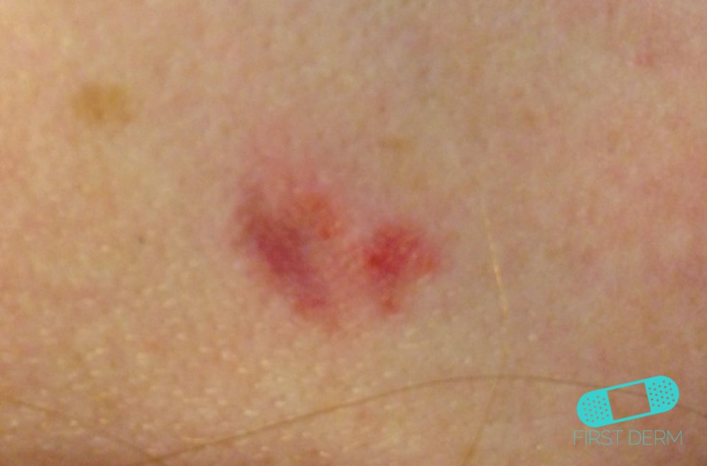
There are four main clinical variants of basal cell carcinoma. These are nodular, superficial spreading, sclerosing and pigmented basal cell carcinomas. Nodular basal cell carcinoma is clinically manifested as a translucent nodule, often with telangiectatic vessels being very evident.
What does basal cell carcinoma mean in medical terms?
Listen to pronunciation. (BAY-sul sel KAR-sih-NOH-muh) Cancer that begins in the lower part of the epidermis (the outer layer of the skin). It may appear as a small white or flesh-colored bump that grows slowly and may bleed.
What are the four major clinicopathologic types of basal cell carcinoma?
Several different clinicopathologic types of BCC exist, each with distinct biologic behavior:Nodular - Cystic, pigmented, keratotic.Infiltrative.Micronodular.Morpheaform.Superficial.
When is Mohs indicated?
Mohs surgery is used to treat the most common skin cancers, basal cell carcinoma and squamous cell carcinoma, as well as some kinds of melanoma and other more unusual skin cancers. Mohs surgery is especially useful for skin cancers that: Have a high risk of recurrence or that have recurred after previous treatment.Aug 18, 2020
Why is it called basal cell carcinoma?
Skin cancer begins in the cells that make up the outer layer (epidermis) of your skin. One type of skin cancer called basal cell carcinoma begins in the basal cells, which make skin cells that continuously push older cells toward the surface.Oct 1, 2021
What is the histologic evidence of malignancy in basal cell carcinoma?
Usually, BCCs are well differentiated and cells appear histologically similar to basal cells of the epidermis. Tumor cells of nodular BCC, sometimes called basalioma cells, typically have large, hyperchromatic, oval nuclei and little cytoplasm. Cells appear uniform, and if present, mitotic figures are usually few.Feb 14, 2022
What is Stage 4 basal cell carcinoma?
Stage 4. The cancer can be any size and may have spread to nearby lymph nodes. It has also spread to areas outside the skin, such as to distant organs like the brain or lungs, or has invaded the skeleton (axial or appendicular) or perineural invasion of skull base.
Is basal cell carcinoma malignant or benign?
Basal cell carcinoma (BCC) is most often a benign form of skin cancer caused by exposure to ultraviolet (UV) light. However, it's the most frequently occurring form of all skin cancers, with more than 3 million people developing BCC in the U.S. every year.Oct 12, 2018
What is the difference between basal cell and squamous cell carcinoma?
Squamous Cell Carcinoma Though this form of skin cancer is not usually life-threatening, one major difference between basal cell and squamous cell cancers is that squamous cell cancer are more likely to grow deeper into the layers of your skin and spread to other parts of the body.
What is the code for a primary malignant neoplasm?
A primary malignant neoplasm that overlaps two or more contiguous (next to each other) sites should be classified to the subcategory/code .8 ('overlapping lesion'), unless the combination is specifically indexed elsewhere.
What chapter is neoplasms classified in?
All neoplasms are classified in this chapter, whether they are functionally active or not. An additional code from Chapter 4 may be used, to identify functional activity associated with any neoplasm. Morphology [Histology] Chapter 2 classifies neoplasms primarily by site (topography), with broad groupings for behavior, malignant, in situ, benign, ...
The ICD code C44 is used to code Merkel-cell carcinoma
Merkel-cell carcinoma is a rare and highly aggressive skin cancer, which, in most cases, is caused by the Merkel cell polyomavirus (MCV) discovered by scientists at the University of Pittsburgh in 2008.
ICD-10-CM Neoplasms Index References for 'C44.41 - Basal cell carcinoma of skin of scalp and neck'
The ICD-10-CM Neoplasms Index links the below-listed medical terms to the ICD code C44.41. Click on any term below to browse the neoplasms index.
Equivalent ICD-9 Code GENERAL EQUIVALENCE MAPPINGS (GEM)
This is the official exact match mapping between ICD9 and ICD10, as provided by the General Equivalency mapping crosswalk. This means that in all cases where the ICD9 code 173.41 was previously used, C44.41 is the appropriate modern ICD10 code.
What is the code for a primary malignant neoplasm?
A primary malignant neoplasm that overlaps two or more contiguous (next to each other) sites should be classified to the subcategory/code .8 ('overlapping lesion'), unless the combination is specifically indexed elsewhere.
What chapter is neoplasms classified in?
All neoplasms are classified in this chapter, whether they are functionally active or not. An additional code from Chapter 4 may be used, to identify functional activity associated with any neoplasm. Morphology [Histology] Chapter 2 classifies neoplasms primarily by site (topography), with broad groupings for behavior, malignant, in situ, benign, ...
Popular Posts:
- 1. icd-10-pcs code for right forearm interstitial myositis
- 2. icd 10 code for chronic intracable moderate to severe pain
- 3. icd 9 code for carotid artery disease
- 4. meridian icd code for jehovah witness
- 5. icd 10 code for chronic right plantar fasciitis
- 6. icd 10 code for hyperhidrosis axilla
- 7. icd 10 code for hand dermatitis
- 8. icd 10 code for sepsis due to gram negative bacilli
- 9. icd 10 code for elevated creatnine kinase
- 10. icd 10 code for old meniscus tear