D22.9
What is the ICD 10 code for excluded nevus?
When a type 2 excludes note appears under a code it is acceptable to use both the code (I78.1) and the excluded code together. blue nevus ( ICD-10-CM Diagnosis Code D22 flammeus nevus ( ICD-10-CM Diagnosis Code Q82.5 hairy nevus ( ICD-10-CM Diagnosis Code D22 melanocytic nevus ( ICD-10-CM Diagnosis Code D22
What is the ICD 10 code for nevus of Ota?
Fibrous papule of the nose Nevus of ota, acquired ICD-10-CM D22.39 is grouped within Diagnostic Related Group (s) (MS-DRG v38.0): 606 Minor skin disorders with mcc
What is the ICD 10 code for non-neoplastic neoplasm?
Nevus, non-neoplastic. I78.1 is a billable/specific ICD-10-CM code that can be used to indicate a diagnosis for reimbursement purposes.
What is the ICD 10 code for neoplasm of unspecified conjunctiva?
2018/2019 ICD-10-CM Diagnosis Code D31.00. Benign neoplasm of unspecified conjunctiva. 2016 2017 2018 2019 Billable/Specific Code. D31.00 is a billable/specific ICD-10-CM code that can be used to indicate a diagnosis for reimbursement purposes.
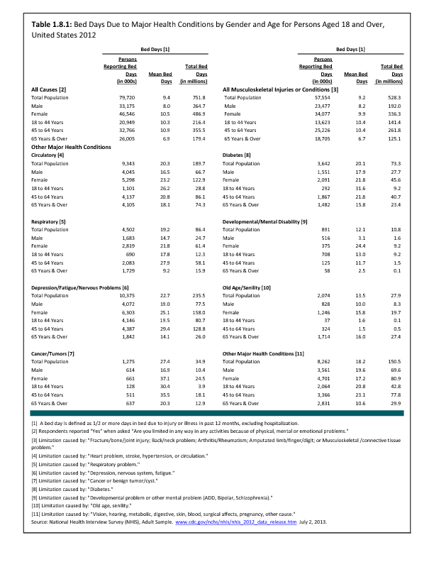
What is the ICD-10 code for melanocytic nevus?
D22.9D22. 9 is a billable/specific ICD-10-CM code that can be used to indicate a diagnosis for reimbursement purposes. The 2022 edition of ICD-10-CM D22.
Is a melanocytic nevus benign?
Melanocytic nevi are benign neoplasms or hamartomas composed of melanocytes, the pigment-producing cells that constitutively colonize the epidermis.
What melanocytic nevus means?
Collapse Section. Giant congenital melanocytic nevus is a skin condition characterized by an abnormally dark, noncancerous skin patch (nevus) that is composed of pigment-producing cells called melanocytes. It is present from birth (congenital) or is noticeable soon after birth.
Is melanocytic nevus a mole?
Moles, also called “melanocytic nevi,” are common in newborns and infants (about 1 percent). If they are seen at birth or develop during the first 1-2 years of life they are called congenital melanocytic nevi. While most of these moles are small, some may be very large.
Is melanocytic nevi melanoma?
Approximately 33% of melanomas are derived directly from benign, melanocytic nevi. Despite this, the vast majority of melanocytic nevi, which typically form as a result of BRAFV600E-activating mutations, will never progress to melanoma.
Is melanocytic melanoma?
Melanocytic neoplasms range from benign lesions, termed melanocytic naevi, to malignant ones, termed melanomas.
How do you say melanocytic nevus?
0:051:02How To Say Melanocytic - YouTubeYouTubeStart of suggested clipEnd of suggested clipMelanocytic or melanocytic melanocytic or melanocytic melanocytic or melanocytic melanocytic orMoreMelanocytic or melanocytic melanocytic or melanocytic melanocytic or melanocytic melanocytic or melanocytic you melanocytic or melanocytic. You melanocytic or melanocytic you.
What is the difference between mole and nevus?
A mole (nevus) is a non-cancerous (benign) skin lesion that is made up of the color-producing (pigment-producing) cells of the skin (melanocytes). A mole that is present at birth is referred to as a congenital nevus. A dysplastic nevus (discussed elsewhere) is a mole in which unusual (atypical) growth is noted.
Does melanocytic mean malignant?
Melanoma is a cancer that begins in the melanocytes. Other names for this cancer include malignant melanoma and cutaneous melanoma. Most melanoma cells still make melanin, so melanoma tumors are usually brown or black. But some melanomas do not make melanin and can appear pink, tan, or even white.
What does melanocytic nevus look like?
They typically appear as small brown, tan, or pink spots. You can be born with moles or develop them later. Moles that you're born with are known as congenital moles.
What is the difference between nevus cells and melanocytes?
Nevus cells are a variant of melanocytes. They are larger than typical melanocytes, do not have dendrites, and have more abundant cytoplasm with coarse granules. They are usually located at the dermoepidermal junction or in the dermis of the skin.
What are the types of nevus?
Blue naevus is a deeply pigmented type of dermal naevus. Cellular naevus is a non-pigmented dermal naevus. Miescher naevus is a dome-shaped smooth dermal naevus often found on the face. Unna naevus is a papillomatous dermal naevus that is in the shape of a raspberry.
What is a Melanocytic Nevi?
A benign (not cancer) growth on the skin that is formed by a cluster of melanocytes (cells that make a substance called melanin, which gives color to skin and eyes). A mole is usually dark and may be raised from the skin.
What is the color of a nevus?
A dysplastic nevus is often larger with borders that are not easy to see. Its color is usually uneven and can range from pink to dark brown. Parts of the mole may be raised above the skin surface. A dysplastic nevus may develop into malignant melanoma (a type of skin cancer).
What is a nevus cell?
The term is usually restricted to nevocytic nevi (round or oval collections of melanin-containing nevus cells occurring at the dermoepidermal junction of the skin or in the dermis proper) or moles, but may be applied to other pigmented nevi. A type of nevus (mole) that looks different from a common mole.
What is a neoplasm composed of melanocytes that usually appears as a dark spot on
A circumscribed stable malformation of the skin and occasionally of the oral mucosa, which is not due to external causes and therefore presumed to be of hereditary origin. A neoplasm composed of melanocytes that usually appears as a dark spot on the skin. A nevus characterised by the presence of excessive pigment.
What is the code for a primary malignant neoplasm?
A primary malignant neoplasm that overlaps two or more contiguous (next to each other) sites should be classified to the subcategory/code .8 ('overlapping lesion'), unless the combination is specifically indexed elsewhere.
What chapter is neoplasms classified in?
All neoplasms are classified in this chapter, whether they are functionally active or not. An additional code from Chapter 4 may be used, to identify functional activity associated with any neoplasm. Morphology [Histology] Chapter 2 classifies neoplasms primarily by site (topography), with broad groupings for behavior, malignant, in situ, benign, ...
What is the table of neoplasms used for?
The Table of Neoplasms should be used to identify the correct topography code. In a few cases, such as for malignant melanoma and certain neuroendocrine tumors, the morphology (histologic type) is included in the category and codes. Primary malignant neoplasms overlapping site boundaries.
What is the code for a primary malignant neoplasm?
A primary malignant neoplasm that overlaps two or more contiguous (next to each other) sites should be classified to the subcategory/code .8 ('overlapping lesion'), unless the combination is specifically indexed elsewhere.
What chapter is neoplasms classified in?
All neoplasms are classified in this chapter, whether they are functionally active or not. An additional code from Chapter 4 may be used, to identify functional activity associated with any neoplasm. Morphology [Histology] Chapter 2 classifies neoplasms primarily by site (topography), with broad groupings for behavior, malignant, in situ, benign, ...
What is the table of neoplasms used for?
The Table of Neoplasms should be used to identify the correct topography code. In a few cases, such as for malignant melanoma and certain neuroendocrine tumors, the morphology (histologic type) is included in the category and codes. Primary malignant neoplasms overlapping site boundaries.
Can multiple neoplasms be coded?
For multiple neoplasms of the same site that are not contiguous, such as tumors in different quadrants of the same breast, codes for each site should be assigned. Malignant neoplasm of ectopic tissue. Malignant neoplasms of ectopic tissue are to be coded to the site mentioned, e.g., ectopic pancreatic malignant neoplasms are coded to pancreas, ...
What is a nevus?
Nevus (or naevus, plural nevi or naevi, from nævus, Latin for "birthmark") is the medical term for sharply-circumscribed [1] and chronic lesions of the skin. These lesions are commonly named birthmarks and moles. Nevi are benign by definition.
Is nevus benign or malignant?
Nevi are benign by definition. Using the term nevus and nevi loosely, most physicians and dermatologists are actually referring to a variant of nevus called the "Melanocytic nevus", which are composed of melanocytes.
Popular Posts:
- 1. icd 9 code for degenerative spine disease
- 2. icd 9 code for pacemaker
- 3. icd 10 code for collapsed bladder
- 4. icd 10 code for neck tension
- 5. icd 10 code for abnormal chest x ray
- 6. icd 10 code for cellulitis of skin on back
- 7. icd 10 code for pink eye right
- 8. icd 10 code for overdose of valium
- 9. icd 10 code for severe right shoulder pain
- 10. icd 10 code for ovarian cysst