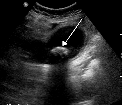
Calcium deposit in bursa, unspecified hand
- M71.449 is a billable/specific ICD-10-CM code that can be used to indicate a diagnosis for reimbursement purposes.
- The 2022 edition of ICD-10-CM M71.449 became effective on October 1, 2021.
- This is the American ICD-10-CM version of M71.449 - other international versions of ICD-10 M71.449 may differ.
Calcium deposit in bursa, unspecified site
M71. 40 is a billable/specific ICD-10-CM code that can be used to indicate a diagnosis for reimbursement purposes. The 2022 edition of ICD-10-CM M71. 40 became effective on October 1, 2021.How many codes in ICD 10?
- ICD-10 codes were developed by the World Health Organization (WHO) External file_external .
- ICD-10-CM codes were developed and are maintained by CDC’s National Center for Health Statistics under authorization by the WHO.
- ICD-10-PCS codes External file_external were developed and are maintained by Centers for Medicare and Medicaid Services. ...
What are the new ICD 10 codes?
The new codes are for describing the infusion of tixagevimab and cilgavimab monoclonal antibody (code XW023X7), and the infusion of other new technology monoclonal antibody (code XW023Y7).
What does ICD 10 mean?
ICD-10 is the 10th revision of the International Statistical Classification of Diseases and Related Health Problems (ICD), a medical classification list by the World Health Organization (WHO). It contains codes for diseases, signs and symptoms, abnormal findings, complaints, social circumstances, and external causes of injury or diseases.
What ICD 10 cm code(s) are reported?
What is the correct ICD-10-CM code to report the External Cause? Your Answer: V80.010S The External cause code is used for each encounter for which the injury or condition is being treated.
What is diagnosis code Z71 89?
Other specified counselingICD-10 code Z71. 89 for Other specified counseling is a medical classification as listed by WHO under the range - Factors influencing health status and contact with health services .
What is DX code R26 81?
Unsteadiness on feetICD-10 code R26. 81 for Unsteadiness on feet is a medical classification as listed by WHO under the range - Symptoms, signs and abnormal clinical and laboratory findings, not elsewhere classified .
What is A41 89?
ICD-10 code A41. 89 for Other specified sepsis is a medical classification as listed by WHO under the range - Certain infectious and parasitic diseases .
What is diagnosis code m89 9?
9: Disorder of bone, unspecified.
What is ICD-10 code for osteoporosis?
0 – Age-Related Osteoporosis without Current Pathological Fracture. ICD-Code M81. 0 is a billable ICD-10 code used for healthcare diagnosis reimbursement of Age-Related Osteoporosis without Current Pathological Fracture.
What is the ICD-10 code for osteoarthritis?
ICD-10 code M19. 90 for Unspecified osteoarthritis, unspecified site is a medical classification as listed by WHO under the range - Arthropathies .
What is the diagnosis for ICD-10 code r50 9?
9: Fever, unspecified.
What is the ICD-10 code for CVA?
I63. 9 - Cerebral infarction, unspecified | ICD-10-CM.
What is the ICD-10-CM code for GPC bacteremia?
ICD-10-CM Code for Bacteremia R78. 81.
What is the ICD-10 code for bone lesion?
Other specified disorders of bone, other site M89. 8X8 is a billable/specific ICD-10-CM code that can be used to indicate a diagnosis for reimbursement purposes. The 2022 edition of ICD-10-CM M89. 8X8 became effective on October 1, 2021.
What is a lytic bone lesion?
Also known as bone lesions or osteolytic lesions, lytic lesions are spots of bone damage that result from cancerous plasma cells building up in your bone marrow. Your bones can't break down and regrow (your doctor may call this remodel) as they should.
What is the correct ICD-10 code for leukocytosis?
288.60 - Leukocytosis, unspecified | ICD-10-CM.
What is a staghorn calculus?
Staghorn calculus. Staghorn calculus (kidney stone) Uric acid nephrolithiasis. Uric acid renal calculus. Clinical Information. A disorder characterized by the formation of crystals in the pelvis of the kidney. A kidney stone is a solid piece of material that forms in the kidney from substances in the urine.
How do you know if you have kidney stones?
The following may be signs of kidney stones that need a doctor's help: extreme pain in your back or side that will not go away. blood in your urine. fever and chills. vomiting. urine that smells bad or looks cloudy.
How do calcium channel blockers help with calcinosis?
Calcium-channel blockers may treat calcinosis cutis by stopping the inward flow of calcium ions into the cells of affected tissues and local macrophages, preventing subsequent crystallization within the mitochondria and cell death.
What is a subepidermal calcified nodule?
Some subtypes of idiopathic calcinosis cutis, however, do have distinct distributions, such as a subepidermal calcified nodule that presents most often as a solitary verrucous nodule on the head or neck of a young child .
What is the differential diagnosis of calcinosis cutis?
The differential diagnosis of calcinosis cutis includes: Milia (which will be differentiated based on the type of material expressed and histopathology) Calciphylaxis (which is more likely to be painful, on the lower extremities, and be associated with livedo mottling and calcification of subcutaneous blood vessels)
What is a metastatic calcinosis cutis?
Metastatic calcinosis cutis presents with altered calcium and phosphorus metabolism, typically in the setting of chronic renal disease. Iatrogenic depositions develop after a medical intervention leads to the inadvertent placement of calcium-containing solutions, such as extravasated intravenous calcium gluconate, within the dermis.
Where is tumoral calcinosis most commonly found?
Tumoral calcinosis presents as large, painless periarticular calcifications, most commonly in the hips and shoulders. Iatrogenic lesions will be located at the site of a recent medical intervention, such as an IV, electrode placement, or repeated heel stick in a neonate.
Can calcinosis cutis cause calcification?
Of the few patients with SLE and calcinosis cutis, most develop calcifications after long-standing SLE. A few cases of calcinosis cutis in the setting of Sjögren’s syndrome have been reported. Benign or malignant neoplasms may also serve as inciting events for dystrophic calcifications.
Is scrotal calcinosis idiopathic?
Scrotal calcinosis has traditionally been classified as a type of idiopathic calcinosis cutis that presents as multiple, firm nodules on the scrotum. However, more recently a close connection between the calcium depositions and epithelial cysts, many with evidence of inflammation, has been demonstrated.

Popular Posts:
- 1. icd 10 code for depression related to chronic disease
- 2. icd 10 code for mass in the kidney
- 3. icd 10 code for dysautonomia
- 4. icd 10 code for stage 2 pressure ulcer mid dorsum
- 5. icd 10 code for uckd unspecified
- 6. icd 10 code for aftercare for end of life care
- 7. icd code for radiculitis
- 8. 2017 icd 10 code for stab wound left upper arm
- 9. icd 10 code for place of occurrence car
- 10. icd 10 code for activity pulling brake release