Full Answer
What is the ICD 10 code for diverticulum with diverticula?
Diagnosis Index entries containing back-references to K22.5: Diverticulum, diverticula (multiple) K57.90 ICD-10-CM Diagnosis Code K57.90 Esophagocele K22.5 Pouch esophagus, esophageal, congenital Q39.6 ICD-10-CM Diagnosis Code Q39.6 Zenker's diverticulum K22.5 (esophagus)
What is the pathophysiology of calyceal diverticula?
The most common theory for the origin of calyceal diverticula is a failure of regression of the third and fourth-generation ureteric buds, as a result of obstructing stones or infection. There are two categories of calyceal diverticula: Only a diverticulum containing milk of calcium will be visible on a plain radiograph.
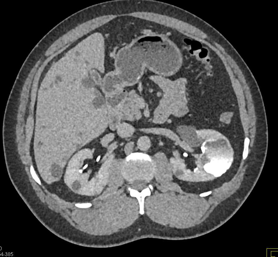
How is a calyceal diverticulum diagnosed on a CT scan?
On nephrographic phase contrast-enhanced CT, a calyceal diverticulum will have an appearance similar to that of a simple cyst. The diagnosis is made with certainty in the excretory phase when the cystic structure fills with contrast material due to communication with the collecting system, and layering of contrast material is seen within.
What is the CPT code for calyceal stone?
Because CPT offers no other specific diagnosis code to describe a calyceal stone, you can use 592.0 for this pathology and for calculi found in any location within the kidney.
What is diagnosis code N28 89?
ICD-10 code N28. 89 for Other specified disorders of kidney and ureter is a medical classification as listed by WHO under the range - Diseases of the genitourinary system .
What is the ICD-10 code for urethral diverticulum?
ICD-10 code: N36. 1 Urethral diverticulum | gesund.bund.de.
What is the ICD-10 code for renal lesion?
The 2022 edition of ICD-10-CM N28. 9 became effective on October 1, 2021. This is the American ICD-10-CM version of N28.
What does diagnosis N20 0 mean?
0: Calculus of kidney.
What causes a urethral diverticulum?
What Causes a Urethral Diverticulum? Urethral diverticulums may be present at birth or develop with time. Often, urethral diverticulum happens in people who have experienced multiple bladder infections, which may weaken the urethral wall. A block of the glands near the urethra may also lead to a urethral diverticulum.
What is bladder diverticulum?
A bladder diverticulum is a pouch in the bladder wall that a person may either be born with ("congenital") or get later ("acquired"). A congenital bladder diverticulum forms when some of the bladder lining pokes through a weak part in the bladder wall.
What is the origin of calyceal diverticula?
The most common theory for the origin of calyceal diverticula is a failure of regression of the third and fourth-generation ureteric buds, as a result of obstructing stones or infection. There are two categories of calyceal diverticula: type I: more common, communicates with a minor calyx.
What is the name of the congenital outpouchings from the renal calyx or pelvis into
Calyceal diverticulum. Calyceal diverticula, also known as pyelocalyceal diverticula are congenital outpouchings from the renal calyx or pelvis into the renal cortex. These diverticula are lined with transitional cell epithelium.
Can a diverticulum be seen on a plain radiograph?
Only a diverticulum containing milk of calcium will be visible on a plain radiograph. It appears as a meniscus-shaped density on an upright radiograph that changes its shape with changing position, i.e. either supine or decubitus radiographs.

Popular Posts:
- 1. icd 9 code for kidney stone with colic
- 2. icd 10 code for suspiciious skin lesion left arm
- 3. icd 10 code for hypermammoplasty
- 4. icd-10 code for charcot in in dm
- 5. icd 9 code for scoloisis
- 6. icd 10 code for cancer piriform sinus
- 7. icd 10 code for multinoduler goiter
- 8. icd 10 code for person waiting on heart transplant candidate
- 9. icd code for edema leg
- 10. icd 10 code for cardiogenic syncope