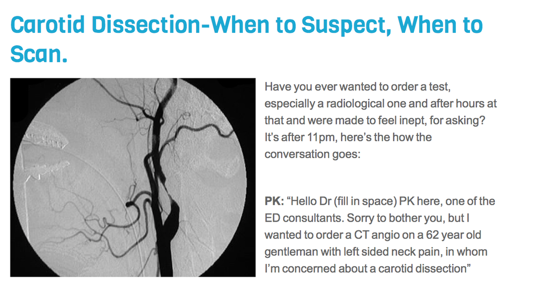
Does carotid artery dissection have a cure?
Surgical or endovascular treatment is more appropriate for symptomatic carotid artery stenosis. Medical treatment. Anticoagulation has traditionally been the treatment of choice for carotid artery dissection. Lately some have suggested treating patients with carotid dissection with antiplatelet medication such as aspirin.
What are the symptoms of a carotid artery aneurysm?
- slight facial drooping
- excessive tiredness or sleeping
- slight muscle weakness in one side of the body
- slurred speech or difficulty speaking
- dizziness
What causes occlusion of the carotid artery?
carotid artery occlusion may be caused by different disease entities, by far the most frequent cause remains atherosclerosis. However, because of uncertainty about the pathophysiology of symptomatic internal carotid artery (ICA) occlusion, there has been contro-versy surrounding its proper management. Natural History of Carotid Artery Occlusion
What is the known cause for carotid artery ulcers?
These are signs and symptoms of a stroke or TIA:
- Sudden numbness or weakness in the face or limbs, often on only one side of the body
- Sudden trouble speaking and understanding
- Sudden vision problems in one or both eyes
- Sudden dizziness or loss of balance
- Sudden, severe headache with no known cause

Where is cavernous carotid artery?
The cavernous segment, or C4, of the internal carotid artery begins at the petrolingual ligament and extends to the proximal dural ring, which is formed by the medial and inferior periosteum of the anterior clinoid process. The cavernous segment is surrounded by the cavernous sinus.
What is a cavernous ICA?
The CCA is a unique aneurysmal lesion because rupture can present in many different forms, namely rupture into the subarachnoid space, into the cavernous sinus proper, and into the surrounding sphenoid sinuses. The risk of CCA rupture is thought to be dependent on multiple factors, most commonly aneurysm size.
Is cavernous carotid intracranial?
The internal carotid arteries (ICA) and their major branches are referred to as internal carotid system. Anatomically the ICA is divided into extracranial and intracranial parts. The intracranial part is further subdivided into petrous, cavernous, and cerebral parts [1,2].
What is the ICD-10 code for carotid cavernous fistula?
I77. 0 is a billable/specific ICD-10-CM code that can be used to indicate a diagnosis for reimbursement purposes. The 2022 edition of ICD-10-CM I77.
What is bilateral cavernous?
Bilateral cavernous carotid aneurysms (CCAs) represent a rare medical condition that can mimic other disorders. We present a rare case of bilateral CCAs simulating an ocular myasthenia.
What is a right cavernous ICA aneurysm?
Aneurysms involving the cavernous segment of the internal carotid artery may produce cranial nerve dysfunction by compression, and occasionally rupture or ischemic events 1,2. The management of symptomatic cavernous aneurysms has evolved over a long period of time.
Is the internal carotid artery an intracranial artery?
The ICA enters the skull base through the carotid canal within the petrous portion of the temporal bone and ascends within the cavernous sinus. It crosses the anterior clinoid process and bifurcates into the ACA and middle cerebral artery (MCA)....Internal Carotid Artery.Anatomical SegmentsBranchesC3-LacerumNo branches6 more rows
What parts are of internal carotid artery?
The Internal Carotid Artery (ICA) is commonly divided into segments: (1) The Cervical segment runs from above the carotid bulb through the neck to the base of the skull; (2) the Petrous segment runs from the base of the skull through the petrous bone; (3) the Cavernous segment runs through the cavernous sinus (note the ...
What is the difference between internal and external carotid artery?
There are two carotid arteries, one on the right and one on the left. In the neck, each carotid artery branches into two divisions: The internal carotid artery supplies blood to the brain. The external carotid artery supplies blood to the face and neck.
What is indirect carotid cavernous fistula?
Indirect carotid-cavernous fistulas are connections between the cavernous sinus and meningeal branches of the external carotid, internal carotid or a combination of both. Indirect carotid-cavernous fistulas most commonly occur spontaneously.
How common is a carotid cavernous fistula?
Carotid cavernous fistulas (CCFs) are a rare but potentially devastating cause of orbital symptoms, visual loss and periocular disfigurement. CCF patients typically present with proptosis, elevated intraocular pressure, prominent tortuous conjunctival vessels and sometimes headache.
What is the ICD-10 code for cavernous malformation?
Q28. 3 - Other malformations of cerebral vessels. ICD-10-CM.
What causes a carotid artery to be damaged?
Damage to the carotid artery. Causes include blunt injuries ( e.g., motor vehicle accidents and sports-related injuries) and penetrating traumas (e.g., gunshot and knife injuries). Damages to the carotid arteries caused either by blunt force or penetrating trauma, such as craniocerebral trauma; thoracic injuries; and neck injuries.
When will the ICD-10-CM S15.0 be released?
The 2022 edition of ICD-10-CM S15.0 became effective on October 1, 2021.
What is the secondary code for Chapter 20?
Use secondary code (s) from Chapter 20, External causes of morbidity, to indicate cause of injury. Codes within the T section that include the external cause do not require an additional external cause code. Type 1 Excludes.
What is the cavernous sinus?
The cavernous sinus (CS) is a paired dural venous sinus centered around the sella turcica. It contains multiple venous channels extending from the endosteal dural layer of the sphenoid bone inferiorly and medially to the meningeal dural layer of the floor of the middle cranial fossa. The CS extends from the superior orbital fissure (SOF) anteriorly to the petrous apex posteriorly, between the dorsum sellae medially and Meckel’s cave laterally. The CS serves as a confluence of venous systems draining the orbit (via superior and inferior ophthalmic veins, SOV, IOV), Sylvian fissure, anterior and middle cranial fossae (via sphenoparietal sinus) and posterior cranial fossa (via the basilar plexus and superior and inferior petrosal sinuses), and they are connected by intercavernous sinuses existing within the sella. The CS also drains inferiorly via emissary veins through the foramen ovale into the pterygoid venous plexus. The venous drainage patterns of the cavernous sinus are pertinent for the pathophysiology of CCFs.
Which segment of the ICA enters the cavernous sinus?
The ICA enters the cavernous sinus as it passes the petrolingual ligament and exits at the proximal dural ring becoming the clinoidal segment.
How to diagnose CCFs?
A complete history and a detailed clinical exam with appropriate diagnostic testing can help identify and classify CCFs. In office modalities that can be performed include auscultation for an ocular bruit and pneumotonometry to assess for increased ocular pulse amplitude. An ocular doppler ultrasound can identify SOV dilation, arterial flow within the SOV, or enlargement of the extraocular muscles. While a specific diagnosis of direct CCFs is often made quickly, the majority of dural CCFs present insidiously and often go months without a diagnosis. The workup may vary significantly given the clinical symptoms, potential etiologies and pathological states associated with CCFs.

Popular Posts:
- 1. icd 10 code for afp screening
- 2. icd 10 code for right heart catheterization
- 3. what is the icd 10 code for syndrome of inappropriate secretion of antidiuretic hormone
- 4. icd 10 code for occlusive thrombosis greater saphenous vein
- 5. icd 10 code for carotid disease
- 6. icd 10 code for fall at home in yard
- 7. icd 10 code for multiple pigmented nevi
- 8. icd 10 cm code for mental health exam
- 9. icd 10 code for myocardial infarction history
- 10. icd 10 code for l wrist fracture