ICD-10 DESCRIPTION ICD-10 DESCRIPTION L83 Acanthosis nigricans D18.01 Hemangioma/cherry angioma L70.0 Acne vulgaris B00.9 Herpes simplex, NOS L70.9 Acne, unspecified L73.2 Hidradenitis suppurativa
D18.01
What is the CPT code for Cherry angioma?
Oct 01, 2021 · 2016 2017 2018 2019 2020 2021 2022 Billable/Specific Code. D18.00 is a billable/specific ICD-10-CM code that can be used to indicate a diagnosis for reimbursement …
What is the ICD 10 code for hemangioma?
ICD-10-CM Diagnosis Code D18.02. Hemangioma of intracranial structures. 2016 2017 2018 2019 2020 2021 2022 Billable/Specific Code. malignant - see Neoplasm, connective tissue, …
What is the ICD 10 code for neoplasm of the trunk?
Oct 01, 2021 · 2022 ICD-10-CM Diagnosis Code D23.5 2022 ICD-10-CM Diagnosis Code D23.5 Other benign neoplasm of skin of trunk 2016 2017 2018 2019 2020 2021 2022 Billable/Specific …
What is the ICD 10 code for malignant melanoma of the trunk?
Oct 01, 2021 · 2016 2017 2018 2019 2020 2021 2022 Billable/Specific Code. L11.1 is a billable/specific ICD-10-CM code that can be used to indicate a diagnosis for reimbursement …
Is Angioma the same as hemangioma?
What is an angioma? Angioma or haemangioma (American spelling 'hemangioma') describes a benign vascular skin lesion. An angioma is due to proliferating endothelial cells; these are the cells that line the inside of a blood vessel.
What is capillary hemangioma?
A benign (not cancer) blood vessel tumor that usually forms on the skin. It may also form on mucous membranes and inside capillaries (small blood vessels) or other places on the body. Lobular capillary hemangiomas usually appear as raised, bright red lesions that may grow quickly and bleed a lot.
What is hemangioma of skin and subcutaneous tissue?
Hemangiomas of the skin can form in the top layer of skin or in the fatty layer underneath, which is called the subcutaneous layer. At first, a hemangioma may appear to be a red birthmark on the skin. Slowly, it will start to protrude upward from the skin. However, hemangiomas are not usually present at birth.
What is hemangioma of skin?
A hemangioma (he-man-jee-O-muh) is a bright red birthmark that shows up at birth or in the first or second week of life. It looks like a rubbery bump and is made up of extra blood vessels in the skin. A hemangioma can occur anywhere on the body, but most commonly appears on the face, scalp, chest or back.Mar 23, 2021
What causes cherry angiomas?
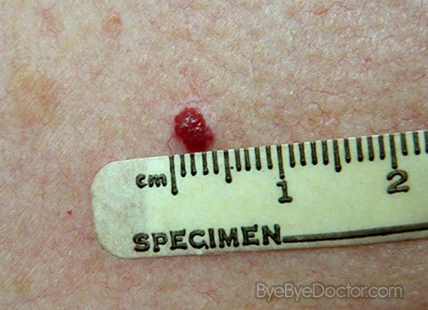
Cherry angiomas are fairly common skin growths that vary in size. They can occur almost anywhere on the body, but usually develop on the trunk. They are most common after age 30. The cause is unknown, but they tend to be inherited (genetic).
What are the two types of hemangiomas?
The two main types of infantile hemangiomas are:
- Superficial hemangiomas, or cutaneous ("in-the-skin") hemangiomas, grow on the skin surface. ...
- Deep hemangiomas grow under the skin, making it bulge, often with a blue or purple tint.
What medical conditions are associated with cherry angiomas?
Eruptions of cherry hemangiomata, glomeruloid hemangiomata, pyogenic granulomas, hypertrichosis lanuginosa, vellous hair cysts, steatocystomas, seborrheic keratoses, acquired ichthyosis, and keratoacanthoma have been associated with hematologic abnormalities and malignancies, including multiple myeloma, Hodgkin ...
What are red blood spots called?
Petechiae are tiny spots of bleeding under the skin. They can be caused by a simple injury, straining or more serious conditions. If you have pinpoint-sized red dots under your skin that spread quickly, or petechiae plus other symptoms, seek medical attention.Jun 29, 2021
What is a benign angioma?
An angioma is a benign growth that consists of small blood vessels. These tumors can be located anywhere on the body. Some of the different types include spider angiomas and cherry angiomas. The cause of most types of angiomas is not known. Cherry angiomas are due to aging and do not have any known significance.
Can a child have a cherry angioma?
A cherry angioma can be a small dot to quite large, fairly common, benign, or non-cancerous bright cherry red or purple, smooth, or raised area, usually featuring a bump or dome shaped clusters of tiny blood vessels on the skin. They tend to occur in older people over 30 years of age, but do occur in children.Aug 30, 2021
Can babies get cherry angiomas?
About Infantile Hemangiomas:
Infantile hemangiomas appear after a baby is born, typically within a month. Roughly 4% to 5% of all infants get them, although they are more common in Caucasians, girls, twins, and preterm or low-birth-weight babies.Dec 24, 2018
Infantile hemangiomas appear after a baby is born, typically within a month. Roughly 4% to 5% of all infants get them, although they are more common in Caucasians, girls, twins, and preterm or low-birth-weight babies.Dec 24, 2018
What is the difference between hematoma and hemangioma?
It is not to be confused with hemangioma, which is an abnormal buildup/growth of blood vessels in the skin or internal organs.
...
...
| Hematoma | |
|---|---|
| Contusion (bruise), a simple form of hematoma. | |
| Specialty | Emergency medicine |
What is the code for a primary malignant neoplasm?
A primary malignant neoplasm that overlaps two or more contiguous (next to each other) sites should be classified to the subcategory/code .8 ('overlapping lesion'), unless the combination is specifically indexed elsewhere.
What is a benign skin lesion?
The majority of cases are congenital. A benign skin lesion consisting of dense, usually elevated masses of dilated blood vessels. A benign tumor of the blood vessels that appears on skin. A benign vascular neoplasm characterized by the formation of capillary-sized or cavernous vascular channels.
What chapter is neoplasms classified in?
All neoplasms are classified in this chapter, whether they are functionally active or not. An additional code from Chapter 4 may be used, to identify functional activity associated with any neoplasm. Morphology [Histology] Chapter 2 classifies neoplasms primarily by site (topography), with broad groupings for behavior, malignant, in situ, benign, ...
What is the code for a primary malignant neoplasm?
A primary malignant neoplasm that overlaps two or more contiguous (next to each other) sites should be classified to the subcategory/code .8 ('overlapping lesion'), unless the combination is specifically indexed elsewhere.
What is the code for a primary malignant neoplasm?
A primary malignant neoplasm that overlaps two or more contiguous (next to each other) sites should be classified to the subcategory/code .8 ('overlapping lesion'), unless the combination is specifically indexed elsewhere.
What chapter is neoplasms classified in?
All neoplasms are classified in this chapter, whether they are functionally active or not. An additional code from Chapter 4 may be used, to identify functional activity associated with any neoplasm. Morphology [Histology] Chapter 2 classifies neoplasms primarily by site (topography), with broad groupings for behavior, malignant, in situ, benign, ...

Popular Posts:
- 1. icd 10 code for screening for specified condition
- 2. guidelines to code admission for dialysis icd 10
- 3. icd 10 code for bacterial pneumonia with sepsis
- 4. 2016 icd 10 code for gallbladder ejection fraction
- 5. icd 10 code for abnormal lung ct
- 6. icd-10 code for sneezing
- 7. icd 10 code for gallstones alcohol
- 8. icd 10 code for abdominal ulcer
- 9. icd 10 code for inability to lose weight
- 10. icd 10 code for positive brca test