D18.01
How do I code a Cherry angioma?
· D18.00 is a billable/specific ICD-10-CM code that can be used to indicate a diagnosis for reimbursement purposes. The 2022 edition of ICD-10-CM D18.00 became effective on October 1, 2021. This is the American ICD-10-CM version of D18.00 - other international versions of ICD-10 D18.00 may differ.
What is the ICD 10 version of haemangioma?
ICD-10-CM Diagnosis Code D18.02. Hemangioma of intracranial structures. 2016 2017 2018 2019 2020 2021 2022 Billable/Specific Code. malignant - see Neoplasm, connective tissue, malignant. plexiform D18.00. ICD-10-CM Diagnosis Code D18.00. Hemangioma unspecified site. 2016 2017 2018 2019 2020 2021 2022 Billable/Specific Code.
Are cherry angiomas common in young people?
Hemangioma unspecified site. Arteriovenous angioma; Benign neoplasm, arteriovenous angioma; Cavernous hemangioma; Cherry angioma; Congenital hemangioma; Glomus tumor; …
Is Angioma the same as hemangioma?
What is an angioma? Angioma or haemangioma (American spelling 'hemangioma') describes a benign vascular skin lesion. An angioma is due to proliferating endothelial cells; these are the cells that line the inside of a blood vessel.
What is hemangioma of skin?
A hemangioma (he-man-jee-O-muh) is a bright red birthmark that shows up at birth or in the first or second week of life. It looks like a rubbery bump and is made up of extra blood vessels in the skin. A hemangioma can occur anywhere on the body, but most commonly appears on the face, scalp, chest or back.
What is hemangioma of skin and subcutaneous tissue?
Hemangiomas of the skin can form in the top layer of skin or in the fatty layer underneath, which is called the subcutaneous layer. At first, a hemangioma may appear to be a red birthmark on the skin. Slowly, it will start to protrude upward from the skin. However, hemangiomas are not usually present at birth.
What is ICD-10 code lentigo simplex?
The ICD code L814 is used to code Lentigo.
What are the two types of hemangiomas?
The two main types of infantile hemangiomas are:Superficial hemangiomas, or cutaneous ("in-the-skin") hemangiomas, grow on the skin surface. ... Deep hemangiomas grow under the skin, making it bulge, often with a blue or purple tint.
What is the cause of cherry angiomas?
Cherry angiomas are fairly common skin growths that vary in size. They can occur almost anywhere on the body, but usually develop on the trunk. They are most common after age 30. The cause is unknown, but they tend to be inherited (genetic).
What medical conditions are cherry angiomas linked to?
Eruptions of cherry hemangiomata, glomeruloid hemangiomata, pyogenic granulomas, hypertrichosis lanuginosa, vellous hair cysts, steatocystomas, seborrheic keratoses, acquired ichthyosis, and keratoacanthoma have been associated with hematologic abnormalities and malignancies, including multiple myeloma, Hodgkin ...
What is the difference between hematoma and hemangioma?
It is not to be confused with hemangioma, which is an abnormal buildup/growth of blood vessels in the skin or internal organs....HematomaContusion (bruise), a simple form of hematoma.SpecialtyEmergency medicine2 more rows
What is a strawberry mole?
A strawberry nevus (hemangioma) is a red birthmark named for its color. This red tinge of skin comes from a collection of blood vessels close to the skin's surface. These birthmarks most commonly occur in young children and infants. Though it's called a birthmark, a strawberry nevus doesn't always appear at birth.
What is diagnosis code Z71 89?
Other specified counselingICD-10 code Z71. 89 for Other specified counseling is a medical classification as listed by WHO under the range - Factors influencing health status and contact with health services .
What is a lentigo simplex?
Lentigo simplex lesions are benign (non-cancerous) lesions that cause no harm. However, their appearance is sometimes similar to melanomas or other cancerous lesions so they need to be examined carefully. Also, the presence or development of multiple lentigines may indicate the presence of associated abnormalities.
What is the ICD-10 code for skin lesion?
ICD-10-CM Code for Disorder of the skin and subcutaneous tissue, unspecified L98. 9.
What is the code for a primary malignant neoplasm?
A primary malignant neoplasm that overlaps two or more contiguous (next to each other) sites should be classified to the subcategory/code .8 ('overlapping lesion'), unless the combination is specifically indexed elsewhere.
When will the ICd 10 D18.00 be released?
The 2022 edition of ICD-10-CM D18.00 became effective on October 1, 2021.
What is vascular anomaly?
A vascular anomaly due to proliferation of blood vessels that forms a tumor-like mass. The common types involve capillaries and veins. It can occur anywhere in the body but is most frequently noticed in the skin and subcutaneous tissue. (from stedman, 27th ed, 2000)
What is a benign vascular neoplasm?
It is characterized by the formation of capillary-sized or cavernous vascular channels. The majority of cases are congenital.
What is the code for a primary malignant neoplasm?
A primary malignant neoplasm that overlaps two or more contiguous (next to each other) sites should be classified to the subcategory/code .8 ('overlapping lesion'), unless the combination is specifically indexed elsewhere.
What is a benign vascular neoplasm characterized by the formation of capillary-sized or
A benign vascular neoplasm characterized by the formation of capillary-sized or cavernous vascular channels. A hemangioma characterized by the presence of cavernous vascular spaces. A vascular anomaly due to proliferation of blood vessels that forms a tumor-like mass.
What is vascular anomaly?
A vascular anomaly that is a collection of tortuous blood vessels and connective tissue. This tumor-like mass with the large vascular space is filled with blood and usually appears as a strawberry-like lesion in the subcutaneous areas of the face, extremities, or other regions of the body including the central nervous system.
What is a benign vascular neoplasm?
It is characterized by the formation of capillary-sized or cavernous vascular channels. The majority of cases are congenital.
Can you use D18.0 for reimbursement?
D18.0 should not be used for reimbursement purposes as there are multiple codes below it that contain a greater level of detail.
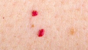
What is a cherry angioma?
Cherry angioma is an asymptomatic firm red, blue, or purple papule, 0.1–1 cm in diameter. When thrombosed, it can appear black in colour until examined with a dermatoscope when the red or purple colour is more easily seen. Cherry angiomas are usually multiple.
How to remove cherry angioma?
If desired for cosmetic reasons, a cherry angioma can be simply removed by one of the following methods: Cryotherapy. Electrosurger y. Vascular laser. See smartphone apps to check your skin.
What genes are involved in cherry angiomas?
Genetic analysis has found cherry angiomas frequently carry specific somatic missense mutations in the GNAQ and GNA11 (Q209H) genes , which are also involved in other vascular and melanocytic proliferations.
Is cherry angioma easy to diagnose?
Cherry angioma is usually easy to diagnose, but occasionally it may be confused with:
Is cherry angioma a malignancy?
There may be a family history of similar lesions. Eruptive cherry angiomas have been rarely reported to be associated with internal malignancy and pregnancy.
Can a cherry angioma be seen in white skin?
Cherry angiomas are very common in males and females of any age or race, with no difference in sexes or races affected. They are however more noticeable in white skin than in skin of colour.
Is cherry angioma a red clod?
Cherry angioma is usually diagnosed clinically and investigation is not necessary for the majority of lesions. It has a characteristic red- clod or lobular pattern on dermoscopy (called lacunar pattern using conventional pattern analysis).

Popular Posts:
- 1. icd 10 code for right scaphoid fracture
- 2. icd 10 code for butcher shop as place of injury
- 3. icd 10 code for depression due to pain
- 4. icd-9-cm code for ecg to monitor a previously diagnosed arrhythmia
- 5. icd 10 cm code for traumatic intracerebral hemorrhage
- 6. icd 10 code for chrst pain
- 7. icd 10 code for left ear itching
- 8. icd-10 code for robbe
- 9. icd 9 code for status post right hip fracture
- 10. icd 10 code for left patella chondromalacia