What ICD-10 code for OCT RNFL?
What is code L98 9?
What is macular thickening?
What is the ICD-10 code for granulation tissue?
What is Z00 01?
What is the ICD-10 code for skin lesion?
What is the difference between macular edema and macular degeneration?
What is eye macula?
What is a macular hole in the eye?
What is granulation tissue?
What is granuloma?
What is the ICD-10 code for granuloma annulare?
How to diagnose cotton wool spots?
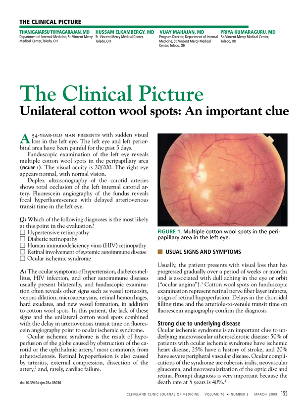
However, fluorescein angiography may reveal areas of capillary nonperfusion adjacent to cotton wool spots . On the other hand, additional work up may be needed to detect the underlying etiology.
What are cotton wool spots?
Signs. On ophthalmic fundus exam, cotton wool spots may appear as small, yellow-white ( or grayish-white), slightly elevated lesions, which look like clouds with a fimbriate border in the superficial retina. Usually they are less than 1/3 disc areas in diameter, and are commonly found in the posterior pole of the funds .
Why are cotton wool spots called cytoid bodies?
They are named cytoid bodies because they look like cells, however they are eosinophilic segments of ganglion cell axons that are swollen because of defective axoplasmic flow. Cytoid bodies are usually packed with accumulations of mitochondria and other intracellular material .
How long does it take for cotton wool spots to disappear?
Work up and treatment are directed towards the underlying etiology. Cotton wool spots classically disappear in 6–12 weeks, however in diabetic retinopathy they may persist for longer .
Can cotton wool spots be asymptomatic?
Cotton wool spots in general are visually asymptomatic, however, a patient may present with vision loss if the fovea is involved . Systemic symptoms of the underlying etiology may be present.
What is a cotton wool spot?
Cotton-wool spots have an ocular differential diagnosis and a systemic differential diagnosis. The ocular differential diagnosis includes the numerous entities that could appear like CWS. The systemic differential diagnosis refers to the underlying conditions that can cause CWS.
How long does a cotton wool spot last?
It has been shown that the size of cotton wool spots in HIV/AIDS are significantly smaller than those in diabetes, hypertension, and CRVO. 13 In addition to these differences, the half-life of CWS in HIV/AIDS is significantly shorter. One study reported the half-life of CWS in AIDS at four to 13 weeks, with a mean of 6.9 weeks. 26 Another study observed a similar time to resolution in hypertensive CWS, which typically resolve in six to 12 weeks. 27

Popular Posts:
- 1. icd 10 code for pulmonary scarring
- 2. icd 10 code for trochanter
- 3. 2016 icd 10 code for abscess knee
- 4. 2015 icd 10 code for dilitation calcis
- 5. icd 10 code for sbo due to adhesions
- 6. icd 10 code for skin assessment
- 7. icd 10 code for viscous supplementation
- 8. 2016 icd 10 code for history of uterine fibroid
- 9. icd 10 code for conjunctivitis left ete
- 10. icd 10 code for m15.4