Aortic ectasia, unspecified site
I77. 819 is a billable/specific ICD-10-CM code that can be used to indicate a diagnosis for reimbursement purposes. The 2022 edition of ICD-10-CM I77. 819 became effective on October 1, 2021.What is the test for aortic root dialation?
There is moderate to severe aortic regurgitation and severe dilatation of his aortic root. A CT scan confirms an ascending aortic aneurysm with a normal aortic arch and descending aorta. A formal ECHO confirms the aortic valve is trileaflet.
How is a dilated aortic root repaired?
Your surgeon will remove the bulging weak area and sew a man-made substitute, called a graft, into place. If the aortic valve is not healthy, your surgeon may repair it or replace it with an artificial valve. After your surgeon does all of the repairs, normal blood flow through your heart and your aorta will resume.
What is the treatment for a mildly dilated aorta?
- Abstract. Evaluate the safety and efficacy of our modified technique of the extravascular procedure for treating mild to moderately dilated ascending aorta in patients with bicuspid aortic valve (BAV) aortopathy.
- Introduction. ...
- Methods. ...
- Results. ...
- Discussion. ...
- Conclusion. ...
- Availability of data and materials. ...
- Abbreviations. ...
- Acknowledgments
- Funding. ...
What is new in dilatation of the ascending aorta?
- Causes of Dilatation of the Ascending Aorta. In patients with aortic dilatation, the aortic wall can be weakened by cystic media degeneration. ...
- Management and Follow-Up in Patients With Aortic Dilatation. ...
- Operative Treatment. ...
- Genetic Counseling. ...
- Conclusions. ...
- Follow-Up of Our Patient. ...
- Disclosures
- Footnotes. ...
What is aortic root dilation?
Otherwise known as an aortic root aneurysm, a dilated aortic root is when the first section of the aorta, where the aortic valve resides, becomes enlarged. When this enlargement reaches a critical size, there is a risk of it rupturing or tearing, leading to a life-threatening situation.
Is aortic root part of thoracic aorta?
The Thoracic Aorta has 4 distinct parts: Aortic Root – Lies in the front portion of the chest below the sternum. It starts at the level of the heart and includes the aortic valve and the portion where the coronary arteries arise called the Sinus of Valsalva.
Where is aortic root located?
The aorta is the large blood vessel that carries blood from the heart to the body. The aortic root is located near where the aorta and the heart connect.
Is aortic dilation and aneurysm the same thing?
Background: The aorta is considered pathologically dilated if the diameters of the ascending aorta and the aortic root exceed the norms for a given age and body size. A 50% increase over the normal diameter is considered aneurysmal dilatation.
What is a mildly dilated aorta?
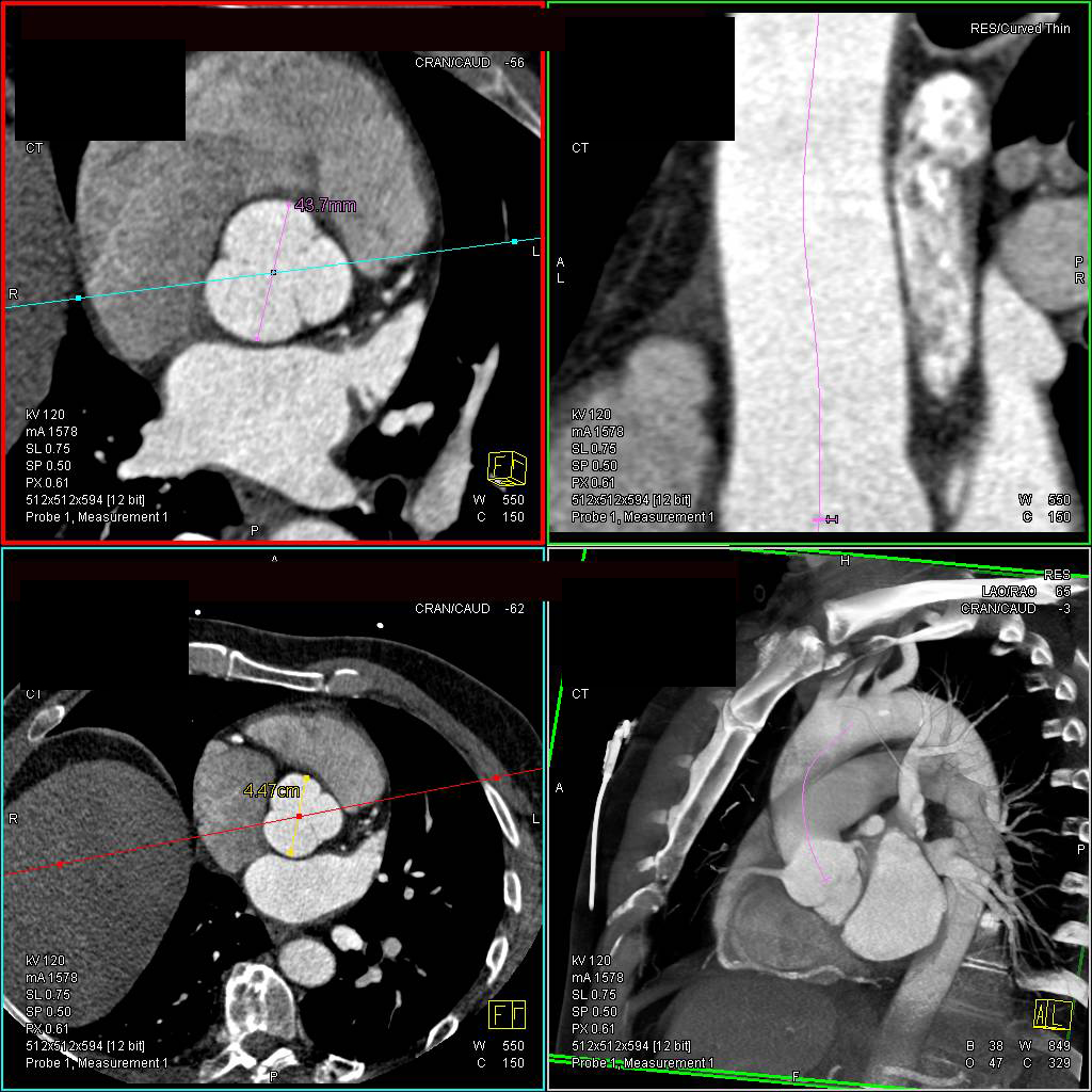
A mild to moderately dilated ascending aorta was defined as having an aorta ascendens dimension between 40 mm to 45 mm on the computer tomography.
Where is an aortic root aneurysm?
An aortic root aneurysm is a bulge in the wall of a specific part of the aorta, your largest artery that carries blood from your heart to the rest of the body. This type of thoracic aortic aneurysm occurs at the point the aorta exits the heart, which is where the aortic valve is located.
Is aortic root the same as aortic valve?
The aortic root is a complex structure that contains the aortic valve and the origins of the left and right coronary arteries. Normal function of the aortic valve can be altered by changes in the size and shape of the aortic root.
Is aortic root the same as aortic sinus?
The spaces between the luminal surface of the three bulges on the aortic root and their respective valvar leaflets are known as the aortic sinuses of Valsalva. Davies considered the wall of the aortic root the aortic sleeve, distinguishing it from the aortic wall on account of its histological compo- sition.
Is aortic root dilation common?
Dilated aortic root (DAR) is a relatively common finding, with a reported prevalence of about 4% measured at the level of the sinuses of Valsalva in the general population.
What is the ICD 10 code for aortic aneurysm?
Abdominal aortic aneurysm, without rupture I71. 4 is a billable/specific ICD-10-CM code that can be used to indicate a diagnosis for reimbursement purposes. The 2022 edition of ICD-10-CM I71. 4 became effective on October 1, 2021.
How common is dilated aorta?
66% of our patients were males and 34% females. 146 patients were found to have aortic dilatation. Therefore, the incidence of aortic dilatation was 6.8% in our study population.
What are the branches of thoracic aorta?
The major noncoronary branches of the thoracic aorta are (in order) the innominate (also known as the brachiocephalic) artery, the left common carotid artery, and the left subclavian artery.
How do you remember the branches of the thoracic aorta?
MnemonicA: arch of aorta.B: brachiocephalic trunk.C: left common carotid artery.S: left subclavian artery.
Is the aortic arch part of the thoracic cavity?
In general, the aortic arch in the thoracic cavity has three primary branches: the brachiocephalic artery (which is divided into the right common carotid artery and right subclavian artery), the left common carotid artery, and the left subclavian artery.
Where does the thoracic aorta end?
Thoracic aorta. The thoracic aorta begins at the level of the T4 vertebra and courses downwards through the posterior mediastinum. Initially, it is found left to the vertebral column, but as it descends it inclines towards the midline and ends up being anterior to the lower border of the body of T12 vertebra.
What is a sac formation?
Clinical Information. A sac formation resulting from the localized dilatation of the wall of the aorta. An abnormal balloon- or sac-like dilatation in the wall of aorta. Most aneurysms -- abnormal bulges or "ballooning" in the wall of an artery -- occur in the aorta.
How is the sac formed?
Sac formed by the dilatation of the wall of the aorta.
Where do abdominal aneurysms occur?
abdominal aortic aneurysms occur in the part of the aorta running through the abdomen.
Which artery carries blood from the heart to the rest of the body?
The aorta is the main artery that carries blood from the heart to the rest of the body. There are two types of aortic aneurysm: thoracic aortic aneurysms occur in the part of the aorta running through the chest. abdominal aortic aneurysms occur in the part of the aorta running through the abdomen.
When will ICD-10-CM I71.9 be released?
The 2022 edition of ICD-10-CM I71.9 became effective on October 1, 2021.
What is the ICd 10 code for aorta dilation?
Congenital dilation of aorta 1 Q25.44 is a billable/specific ICD-10-CM code that can be used to indicate a diagnosis for reimbursement purposes. 2 The 2021 edition of ICD-10-CM Q25.44 became effective on October 1, 2020. 3 This is the American ICD-10-CM version of Q25.44 - other international versions of ICD-10 Q25.44 may differ.
When will the ICD-10-CM Q25.44 be released?
The 2022 edition of ICD-10-CM Q25.44 became effective on October 1, 2021.
What is the ICd 10 code for aorta malformation?
Other congenital malformations of aorta 1 Q25.4 should not be used for reimbursement purposes as there are multiple codes below it that contain a greater level of detail. 2 The 2021 edition of ICD-10-CM Q25.4 became effective on October 1, 2020. 3 This is the American ICD-10-CM version of Q25.4 - other international versions of ICD-10 Q25.4 may differ.
What does "exclude note" mean?
A type 1 excludes note is a pure excludes. It means "not coded here". A type 1 excludes note indicates that the code excluded should never be used at the same time as Q25.4. A type 1 excludes note is for used for when two conditions cannot occur together, such as a congenital form versus an acquired form of the same condition.
When will the ICD-10-CM Q25.4 be released?
The 2022 edition of ICD-10-CM Q25.4 became effective on October 1, 2021.
What is the A00-B99?
certain conditions originating in the perinatal period ( P04 - P96) certain infectious and parasitic diseases ( A00-B99) complications of pregnancy, childbirth and the puerperium ( O00-O9A)
When will ICD-10-CM I71.2 be released?
The 2022 edition of ICD-10-CM I71.2 became effective on October 1, 2021.

Popular Posts:
- 1. icd 9 code for mole flank
- 2. icd 10 code needed for a1c
- 3. icd 10 cm code for pancreatitis
- 4. icd 10 code for history of aki
- 5. icd 9 code for acute febrile illness
- 6. icd-9-cm code for late effects speech deficit due to cva cause
- 7. icd 9 code for uretrovesiculorectal fistula
- 8. icd 10 code for myositis
- 9. icd-10 dx code for ild
- 10. icd 10 code for tpn management