Retinal edema
- H35.81 is a billable/specific ICD-10-CM code that can be used to indicate a diagnosis for reimbursement purposes.
- The 2022 edition of ICD-10-CM H35.81 became effective on October 1, 2021.
- This is the American ICD-10-CM version of H35.81 - other international versions of ICD-10 H35.81 may differ.
Puckering of macula, unspecified eye
The 2022 edition of ICD-10-CM H35. 379 became effective on October 1, 2021.What are the new ICD 10 codes?
The new codes are for describing the infusion of tixagevimab and cilgavimab monoclonal antibody (code XW023X7), and the infusion of other new technology monoclonal antibody (code XW023Y7).
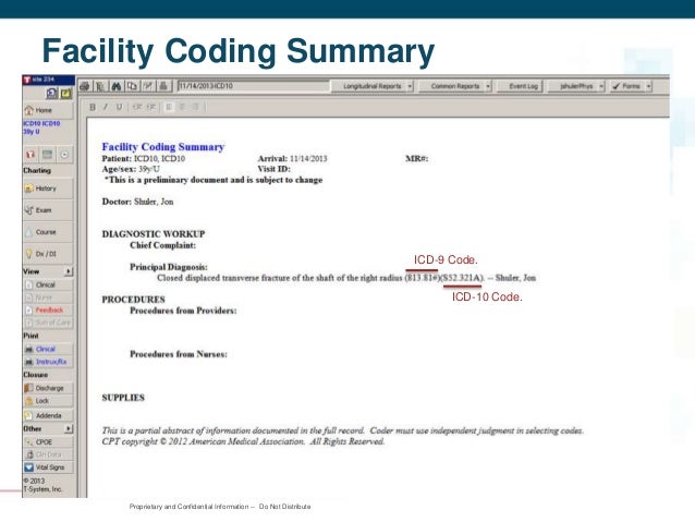
What are the new features of ICD 10?
- ICD-10-CM consists of 21 chapters.
- Some chapters include the addition of a sixth character.
- ICD-10-CM includes full code titles for all codes (no references back to common fourth and fifth digits).
- V and E codes are no longer supplemental classifications.
- Sense organs have been separated from nervous system disorders.
Where can one find ICD 10 diagnosis codes?
Search the full ICD-10 catalog by:
- Code
- Code Descriptions
- Clinical Terms or Synonyms
What is the ICD 10 code for surgery clearance?
A preoperative examination to clear the patient for surgery is part of the global surgical package, and should not be reported separately. You should report the appropriate ICD-10 code for preoperative clearance (i.e., Z01. 810 – Z01. 818) and the appropriate ICD-10 code for the condition that prompted surgery.

What is the ICD-10 code for epiretinal membrane left eye?
Macular pucker occurs when a contracting epiretinal membrane distorts the underlying retina.
What is ERM of the eye?
Epiretinal membrane (ERM) is a condition where a sheet of naturally occurring cells develops on or above the surface of the central part of your retina, an area called the macula. ERM can affect vision if this sheet of cells starts to shrink, causing the retina to wrinkle up under it.
How do you code an epiretinal membrane?
Disease Entity. Macular Pucker ICD-9 code 362.56. Numerous terms have been used to describe this entity including: Epiretinal membrane, epimacular membrane, surface-wrinkling retinopathy, cellophane maculopathy, and preretinal macular fibrosis.
Is ERM and macular pucker the same?
Macular Pucker, also known as an Epiretinal Membrane (ERM) is an eye condition that affects the macula, the sweet spot of center vision. The back of your eye is lined by the retina, the light seeing layer in the back of the eye.
When do you use ERM?
Patients with moderate visual loss, recent onset of symptoms, or progression are the best candidates for ERM surgery. Functional outcome in patients with poor initial visual acuity or long-standing disease is unsatisfactory.
Is epiretinal membrane a retinopathy?
An epiretinal membrane is also sometimes called a macular pucker, macular fibrosis, surface wrinkling retinopathy or cellophane maculopathy.
What is the ICD-10 code for right eye epiretinal membrane?
Puckering of macula, right eye H35. 371 is a billable/specific ICD-10-CM code that can be used to indicate a diagnosis for reimbursement purposes. The 2022 edition of ICD-10-CM H35. 371 became effective on October 1, 2021.
What causes ERM?
The most common cause of ERM is an age-related condition called posterior vitreous detachment (PVD). In these instances, the vitreous gel filling the eye separates from the retina resulting in micro-tears and symptoms of floaters and flashers.
Coding for Laterality in AMD
When you use the codes for dry AMD (H35.31xx) and wet AMD (H35.32xx), you must use the sixth character to indicate laterality as follows:
Coding for Staging in Dry AMD
The codes for dry AMD—H35.31xx—use the seventh character to indicate staging as follows:
Defining Geographic Atrophy
When is the retina considered atrophic? The Academy Preferred Practice Pattern1 defines GA as follows:
Coding for Geographic Atrophy
The Academy recommends that when coding, you indicate whether the GA involves the center of the fovea: Code H35.31x4 if it does and H35.31x3 if it doesn’t, with “x” indicating laterality.
Coding for Staging in Wet AMD
The codes for wet AMD—H35.32xx—use the sixth character to indicate laterality and the seventh character to indicate staging as follows:
Focus on Payment Policy at AAO 2017
Introduction to Physician Payment Policy (Sym12). A panel will explain how new CPT codes are created and valued; how existing codes are targeted for reevaluation; the impact of new technology on the valuation of existing procedures; and the difference between CMS and commercial carrier coverage policies. When: Sunday, Nov. 12, 11:15 a.m.-12:15 p.m.
What is idiopathic ERM?
Idiopathic ERMs affect the architecture of the macula. There can be blunting of the foveal contour or wrinkling on the retinal surface from membrane contracture. Most commonly it involves the foveal and parafoveal area. Macular edema and/or pseudohole can be seen in association with an ERM.
What instruments are used to remove ERM?
A number of different instruments can be used to facilitate removal including intraocular forceps, pick, diamond dusted instruments , as well as other instruments. In many cases. internal limiting membrane peel is also performed concurrent with ERM iremoval.
What is OCT in medical terms?
This is a clinical diagnosis based on history and clinical exam, including slit lamp and dilated fundus examination. In some cases, Optical Coherence Tomography (OCT) is useful in the diagnosis, quantification of retinal thickness, and management of this condition.
Where is the epiretinal membrane located?
An epiretinal membrane (ERM) is a fibrocellular tissue found on the inner surface of the retina. It is semi-translucent and proliferates on the surface of the internal limiting membrane.
Is ERM bilateral or bilateral?
Careful examination of the fellow eye is also recommended given that ERMs are bilateral in approximately 10-20% of patients.
Popular Posts:
- 1. icd 10 code for bone infarct
- 2. icd 10 code for prolonged qt.
- 3. icd 10 code for bilateral hand cellulitis
- 4. icd 10 code for dementia with sundowning
- 5. icd-10-cm code for pure hypercholesterolemia
- 6. icd 10 code for status right shoulder tendinitis
- 7. icd 10 code for left foot osteopenia
- 8. icd 10 code for end stage glenohumeral arthritis
- 9. icd 10 code for pathological left femur fracture
- 10. icd 10 diagnosis code for liver mass