Hemangioma of intra-abdominal structures
D18. 03 is a billable/specific ICD-10-CM code that can be used to indicate a diagnosis for reimbursement purposes. The 2022 edition of ICD-10-CM D18. 03 became effective on October 1, 2021.What is the ICD 10 code for hemangioma?
Hemangioma of other sites. D18.09 is a billable/specific ICD-10-CM code that can be used to indicate a diagnosis for reimbursement purposes. The 2019 edition of ICD-10-CM D18.09 became effective on October 1, 2018. This is the American ICD-10-CM version of D18.09 - other international versions of ICD-10 D18.09 may differ.
What is the ICD 10 code for neoplasm of esophagus?
2018/2019 ICD-10-CM Diagnosis Code C15.9. Malignant neoplasm of esophagus, unspecified. 2016 2017 2018 2019 Billable/Specific Code. C15.9 is a billable/specific ICD-10-CM code that can be used to indicate a diagnosis for reimbursement purposes.
What is the ICD 10 code for esophageal varices without bleeding?
Esophageal varices without bleeding. I85.00 is a billable/specific ICD-10-CM code that can be used to indicate a diagnosis for reimbursement purposes. The 2019 edition of ICD-10-CM I85.00 became effective on October 1, 2018.
What is esophageal hemorrhage ICD 10?
Esophageal bleeding; Esophageal hemorrhage; Clinical Information. A disorder characterized by bleeding from the esophagus. Bleeding originating from the esophagus. ICD-10-CM K22.8 is grouped within Diagnostic Related Group(s) (MS-DRG v 38.0): 391 Esophagitis, gastroenteritis and miscellaneous digestive disorders with mcc
What is the ICD-10 code for hemangioma?
D18.0ICD-10 code D18. 0 for Hemangioma is a medical classification as listed by WHO under the range - Neoplasms .
What is a hemangioma tumor?
A hemangioma (he-man-jee-O-muh) is a bright red birthmark that shows up at birth or in the first or second week of life. It looks like a rubbery bump and is made up of extra blood vessels in the skin. A hemangioma can occur anywhere on the body, but most commonly appears on the face, scalp, chest or back.
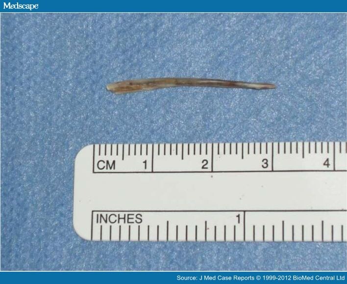
What is hemangioma of intra abdominal structures?
They are benign tumours that arise from embryonic remnants of unipotent angioblastic cells [1]. Although hemangiomas may occur anywhere within the abdomen, including the solid organs, hollow viscera, ligaments, and abdominal wall, the liver is the most common site.
What is the ICD-10-CM code for a cavernous hemangioma?
Other malformations of cerebral vessels The 2022 edition of ICD-10-CM Q28. 3 became effective on October 1, 2021.
What is an internal hemangioma?
An internal hemangioma is a type of noncancerous tumor that forms from the abnormal growth of excess blood vessels. Hemangiomas usually occur on the skin of infants, presenting as a red mark. However, they occasionally develop in internal organs, including the brain and liver.
Is a hemangioma a lesion?
Liver hemangiomas are the most common type of benign liver lesions. They're made up of tangled clumps of blood vessels. Most don't cause symptoms and don't need to be removed.
How many types of hemangiomas are there?
There are three main types: Superficial (on the surface of the skin): These look flat at first, and then become bright red with a raised, uneven surface. Deep (under the skin): These appear as a bluish-purple swelling with a smooth surface. Mixed: These hemangiomas have both superficial and deep components.
What is the cause of hemangiomas?
Hemangiomas of the skin develop when there's an abnormal proliferation of blood vessels in one area of the body. Experts aren't sure why blood vessels group together like this, but they believe it's caused by certain proteins produced in the placenta during gestation (the time when you're in the womb).
What causes a hemangioma in adults?
The cause of hemangiomas and vascular malformations often isn't known. They may be passed on (inherited) in some families. The way they're passed on is called autosomal dominant inheritance. This means that only 1 parent needs to have the gene to pass it on.
Can B96 81 be used as a primary diagnosis?
The note in ICD-10 under codes B95-B97 states that 'these categories are provided for use as supplementary or additional codes to identify the infectious agent(s) in disease classified elsewhere', so you would not use B96. 81 as a primary diagnosis, but as an additional code with the disease listed first.
Can F07 81 be used as a primary diagnosis?
Our physicians have used IDC-10 code F07. 81 as the primary diagnosis for patients presenting with post concussion syndrome.
Can E78 2 and E29 1 be billed together?
For example, E78. 2 Mixed hyperlipidemia cannot be coded with 5-alpha-reductase deficiency (E29. 1 Testicular hypofunction), but the note for this is not at E78.
What is a benign skin lesion?
The majority of cases are congenital. A benign skin lesion consisting of dense, usually elevated masses of dilated blood vessels. A benign tumor of the blood vessels that appears on skin. A benign vascular neoplasm characterized by the formation of capillary-sized or cavernous vascular channels.
Is morphology included in the category and codes?
In a few cases, such as for malignant melanoma and certain neuroendocrine tumors, the morphology (histologic type) is included in the category and codes. Primary malignant neoplasms overlapping site boundaries.
What is the name of the cancer of the esophagus?
Malignant neoplasm of esophagus. Approximate Synonyms. Adenocarcinoma of esophagus. Cancer of the esophagus. Cancer of the esophagus, adenocarcinoma. Cancer of the esophagus, squamous cell. Esophageal cancer metastatic to unspecified site. Metastasis from malignant tumor of esophagus.
What is metastatic esophagus?
Squamous cell carcinoma of esophagus. Clinical Information. A primary or metastatic malignant neoplasm involving the esophagus. The esophagus is a hollow tube that carries food and liquids from your throat to your stomach.
What is the code for a primary malignant neoplasm?
A primary malignant neoplasm that overlaps two or more contiguous (next to each other) sites should be classified to the subcategory/code .8 ('overlapping lesion'), unless the combination is specifically indexed elsewhere.

Popular Posts:
- 1. icd 10 code for chronic lymphedema bilateral lower extremities
- 2. icd 10 code for toe pain left
- 3. icd 10 code for heavy alcohol consumption
- 4. icd 10 code for hiv encephalopathy
- 5. icd-10-cm code for pregnancy state
- 6. icd 10 code for wax impaction
- 7. eye left icd 10 cm code for impacted hearing
- 8. icd 10 code for puncture wound left palm
- 9. icd code for eschar
- 10. icd 9 code for partial tear knee