Other vitreous opacities, unspecified eye
H43. 399 is a billable/specific ICD-10-CM code that can be used to indicate a diagnosis for reimbursement purposes. The 2022 edition of ICD-10-CM H43. 399 became effective on October 1, 2021.What is the best treatment for Eye floaters?
These help:
- Eating antioxidant-rich foods
- Drinking antioxidant-rich juices
- Taking supplements with Taurine (be careful!)
- Taking Omega-3 fatty acids
- Taking serrapeptase enzyme
What does it mean if you have a floater in your eye?
Floaters and spots that appear out of the blue could mean that the retina itself is becoming dislodged from your eye. Floaters and spots typically appear when tiny pieces of the eye's gel-like vitreous break loose within the inner back portion of the eye. Eye floaters can be clumpy or stringy; light or dark.
How to get rid of floaters in the eye?
Quick fixes are:
- drink plenty of water
- get enough good sleep
- eat organically
- take high-quality supplements (including bilberry)
- cut down on alcohol and coffee.
Is there any way of getting rid of Eye floaters?
Try the following:
- Eat a healthy diet full of anti-inflammatory foods.
- Apply hot and cold compresses to help your eyes relax.
- Gently massage your temples with your eyes closed.
- Do eye exercises, such as rolling your eyes and focusing on a moving object, to build resistance to fatigue and reduce floaters.
- Reduce screen time.

What is the code for vitreous floaters right eye?
The 2022 edition of ICD-10-CM H43. 39 became effective on October 1, 2021.
What is ICD-10 code for eye problem?
H57. 9 - Unspecified disorder of eye and adnexa. ICD-10-CM.
What is the ICD-10 code for vitreous degeneration?
ICD-10 Code for Vitreous degeneration, bilateral- H43. 813- Codify by AAPC.
What is the ICD-10 code for vitreous detachment?
CASE 2 – POSTERIOR VITREOUS DETACHMENT (PVD) What ICD-10 code(s) should be used There are two valid diagnoses: H43. 811 (Vitreous degeneration, right eye) and Z96. 1 (Presence of intraocular lens; pseudophakia).
What is a visual disturbance?
Visual disturbance is when you experience a short spell of flashing or shimmering of light in your sight. The symptoms normally last around twenty minutes before your sight returns to normal. Usually, there is no headache during the visual disturbance.
What is the ICD-10 code for chronic dry eyes?
ICD-10-CM Code for Dry eye syndrome H04. 12.
What is the ICD-10 code for PVD?
ICD-10 code I73. 9 for Peripheral vascular disease, unspecified is a medical classification as listed by WHO under the range - Diseases of the circulatory system .
What is the ICD-10-CM code for posterior vitreous detachment?
Vitreous degeneration, unspecified eye H43. 819 is a billable/specific ICD-10-CM code that can be used to indicate a diagnosis for reimbursement purposes. The 2022 edition of ICD-10-CM H43. 819 became effective on October 1, 2021.
What is posterior vitreous detachment?
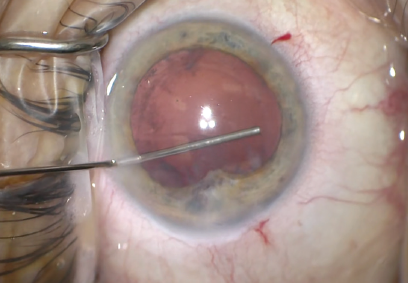
Posterior vitreous detachment (PVD) occurs when the gel that fills the eyeball separates from the retina. It's a natural, normal part of aging. PVD can cause floaters or flashes in your sight, which usually become less noticeable over time. The condition isn't painful, and it doesn't cause vision loss on its own.
How is posterior vitreous detachment diagnosis?
Posterior vitreous detachment is usually diagnosed with a dilated eye examination. However, if the vitreous gel is very clear, it may be hard to see the PVD without additional testing, such as optical coherence tomography (OCT) or ocular ultrasound (see Figure 2).
What happens when the vitreous separates from the retina?
When your vitreous detaches, strands of the vitreous often cast new shadows on your retina — and those shadows appear as floaters. You may also notice flashes of light in your side (peripheral) vision. Sometimes, vitreous detachment causes more serious eye problems that need treatment right away.
What does vitreous degeneration bilateral mean?
During adulthood, the vitreous humor that fills the eye becomes liquid and condenses as the fibers shrink and cause condensed vitreous material. Vitreous degeneration results in dark specks, floaters seen as small moving dots or wispy dark spots or lines, or flashing lights.
What is a floater in the eye?
Floaters are deposits of various size, shape, consistency, refractive index, and motility within the eye's vitreous humour, which is normally transparent. At a young age, the vitreous is transparent, but as one ages, imperfections gradually develop. The common type of floater, which is present in most persons' eyes, ...
Why do people have floaters in their eyes?
The common type of floater, which is present in most persons' eyes, is due to degenerative changes of the vitreous humour. The perception of floaters is known as myodesopsia, or less commonly as myodaeopsia, myiodeopsia, myiodesopsia.
What is the ICD code for vitreous body?
H43.9 is a billable ICD code used to specify a diagnosis of unspecified disorder of vitreous body. A 'billable code' is detailed enough to be used to specify a medical diagnosis.
Why are floaters visible?
Floaters are visible because of the shadows they cast on the retina or refraction of the light that passes through them, and can appear alone or together with several others in one's visual field.
What is the approximate match between ICd9 and ICd10?
This means that while there is no exact mapping between this ICD10 code H43.9 and a single ICD9 code, 379.29 is an approximate match for comparison and conversion purposes.
What is the least appropriate code for uveitis?
The least appropriate code is unspecified. Only use unspecified when there is not a more definitive code. Reviewing the principles of ICD-10 and the classifications of uveitis will help ensure correct ...
What is the best ICD-10 code?
When selecting the appropriate ICD-10, you should choose the code that accurately reflects the initial confirmed diagnosis. The best code is the actual disease. Without a confirmed diagnosis, the next best is a sign or symptom. After that, other is the best option. The least appropriate code is unspecified.
What is the diagnosis of anterior uveitis?
The process of diagnosing anterior uveitis and determining the most specific code is outlined in Figure 1. The initial diagnosis of anterior uveitis (primary acute, recurrent acute, and chronic) is used when waiting for a confirmed diagnosis.
When to use unspecified code?
The least appropriate code is unspecified. Only use unspecified when there is not a more definitive code. Code the diagnosis you know. Do not code probable, suspected, or questionable diagnoses, do not you rule out conditions until they are confirmed. These principles are relevant when coding for uveitis cases.
Is uveitis anterior or posterior?
Based on the anatomical involvement, uveitis can be classified as anterior, affecting the anterior chamber/iris; intermediate, affecting the vitreous/pars plana; posterior, affecting the retina and choroid; or panuveitis, affecting the anterior chamber, vitreous, and retina/choroid.
Popular Posts:
- 1. icd 10 code for forign body of throat
- 2. icd 10 code for small airway disease
- 3. icd 9 code for injury to eye with grinder
- 4. icd 10 code for total bilirubin elevated
- 5. icd 10 code for left sided headache
- 6. icd 10 code for occlusion of left popliteal artery
- 7. icd 10 code for acute kidney injury due to hypovolemia
- 8. icd-10-cm code for borderline cephalopelvic disproportion with obstructed labor
- 9. icd 9 code for cameron lesion
- 10. icd 9 code for buttock contusion