Unspecified fracture of shaft of humerus, right arm, initial encounter for closed fracture. S42. 301A is a billable/specific ICD-10-CM code that can be used to indicate a diagnosis for reimbursement purposes. The 2022 edition of ICD-10-CM S42.
Full
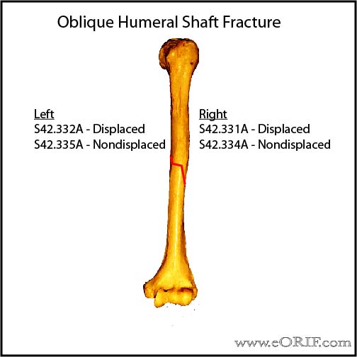
AnswerWhat is the ICD 10 code for right humerus fracture?
Right humerus (upper arm bone) fracture ICD-10-CM S42.401A is grouped within Diagnostic Related Group (s) (MS-DRG v38.0): 562 Fracture, sprain, strain and dislocation except femur, hip, pelvis and thigh with mcc 563 Fracture, sprain, strain and dislocation except femur, hip, pelvis and thigh without mcc
What is humerus fracture?
A traumatic or pathologic injury to the humerus in which the continuity of the humerus is broken.
What is the ICD 10 code for hip fracture?
563 Fracture, sprain, strain and dislocation except femur, hip, pelvis and thigh without mcc Reimbursement claims with a date of service on or after October 1, 2015 require the use of ICD-10-CM codes.
What is the ICD 10 code for upper arm fracture?
ICD-10-CM Codes › S00-T88 Injury, poisoning and certain other consequences of external causes › S40-S49 Injuries to the shoulder and upper arm › S42-Fracture of shoulder and upper arm › Fracture of upper end of humerus S42.2 Fracture of upper end of humerus S42.2-
What is ICD-10 code for fracture of distal humerus?
ICD-10-CM Code for Fracture of lower end of humerus S42. 4.
What is the ICD-10 code for fracture of right humerus?
ICD-10 Code for Unspecified fracture of shaft of humerus, right arm, initial encounter for closed fracture- S42. 301A- Codify by AAPC.
Where is the distal humerus?
The distal humerus is the lower end of the humerus. It forms the upper part of the elbow and makes it possible for your forearm to bend and straighten. The radial head is the knobby end of the radius where it meets the elbow.
What is the ICD-10 code for humeral head fracture?
ICD-10 Code for Fracture of upper end of humerus- S42. 2- Codify by AAPC.
What is the ICD 9 code for fracture of humerus?
79.31 Open reduction of fracture with internal fixation; humerus - ICD-9-CM Vol. 3 Procedure Codes.
What is the right proximal humerus?
The proximal humerus consists of the humeral head, anatomical neck, greater tuberosity, lesser tuberosity, surgical neck, and proximal shaft. Fractures of the proximal humerus (Box 4-8) are associated with osteoporosis. The majority of fractures are the result of indirect forces such as a fall onto an outstretched arm.
Which of the following is distal to the humerus?
Also, the humerus has distal articulations with the radius and ulna at the elbow joint.
What is the classification of this distal humerus fracture?
Distal Humerus FracturesAO/OTA Classification of Distal Humerus FracturesType AExtra-articular (supracondylar fracture), 80% are extension type; epicondyleType BIntraarticular- Single column (partial articular-isolated condylar, coronal shear, epicondyle with articular extension)2 more rows•May 6, 2022
What is an intra articular distal humerus fracture?
Intra-articular fractures of the distal humerus are complex injuries that can considerably limit elbow function if not treated appropriately. Surgical management is indicated for most intra-articular distal humerus fractures with the goal of restoring elbow range of motion and function.
What is a fracture of the humerus?
A humerus fracture is the medical name for breaking the bone in your upper arm (your humerus). Humerus fractures are usually caused by traumas like car accidents or falls. If you break your humerus, you might need surgery to repair your bone.
What is a 3 part proximal humerus fracture?
3-PART FRACTURE: This is when the proximal humerus is broken into three pieces, and there are then two fracture lines on x-ray. This most often involves the greater tuberosity and the surgical neck of the humerus.
What is the humeral head?
The shoulder is considered a ball-and-socket joint with the ball being the rounded end of the humerus (humeral head) and the socket being the cup part of the scapula (glenoid).
What is the ICd 9 code for a fracture of the distal femur?
So a physeal fracture of the distal femur would be reported as 821.22 for a closed fracture or 821.32 for an open fracture. It should be noted that these codes are not specific to Salter-Harris fractures. These codes are used for any fracture or separation of the epiphysis in the lower end of the femur. These codes are reported both for adults (who have closed growth plates) and children and adolescents (who have open growth plates) even though the potential for complications, including arrested bone growth, is much greater for children and adolescents.
What happens when a physeal fracture occurs?
When a physeal fracture occurs, the cartilaginous tissue of the growth plate becomes disrupted or separated, and when this occurs, bone growth may be affected. In the United States, physeal fractures are classified by severity using a system developed in 1963 by Robert Salter and W. Robert Harris; the system is known as ...
What is a growth plate fracture?
Physeal fractures, also referred to as growth plate fractures, are fractures that occur in the distal or proximal physis of the long bones, and they are of particular concern when they occur in children and adolescents who have not finished growing. Until full growth is attained, the growth plates are open and filled with cartilaginous tissue.
How many epiphyses are there in a long bone?
Epiphysis: Each long bone has two epiphyses, one located at the proximal end and one at the distal end. The epiphyses are composed of spongy bone that contains bone marrow. Physis: The physis also is referred to as the growth plate, epiphyseal plate or epiphyseal cartilage.
What is the only site that could be considered a metadiphyseal fracture?
In the proximal radius, the only site that could be considered a "Metadiphyseal Fracture" is the Radial Neck. It would be far better to not use this term for this fracture, and just call it a Radial Neck Fracture, particularly to avoid confusion in coding. You must log in or register to reply here.
What is proximal metadiaphysis?
Metadiaphysis is the joining area of metaphysis and diaphysis region which occur at shaft.
Which part of the bone is the end of the metaphysis?
AlanPechacek. The Metaphysis is the end of a long bone where the growth in the length of the bone occurs in growing children, or occurred in adults, and can be proximal or distal. The Diaphysis is the shaft portion of the long bone, and it does not contribute to growth in length of the bone in growing children or adults.
Which part of the bone does not contribute to growth in length of the bone in growing children or adults?
The Diaphysis is the shaft portion of the long bone, and it does not contribute to growth in length of the bone in growing children or adults. The "Metadiaphysis" is the junction of/between the Metaphysis and the Diaphysis, including growing children and adults.
Popular Posts:
- 1. icd 10 code for right breast infiltrating ductal carcinoma
- 2. icd 10 code for critical limb ischemia right lower extremity
- 3. icd 9 code for new onset type 2 diabetes
- 4. icd 10 code for posterior labral tear shoulder
- 5. icd 10 code for personal history of angioedema
- 6. icd 10 code for secondary thrombocytopenia due to hypersplenism
- 7. icd 10 code for major depressive disorder recurrent mild
- 8. icd 10 code for erythromelalgia
- 9. icd 10 code for pump catheter leak
- 10. icd 10 code for balance difficulties