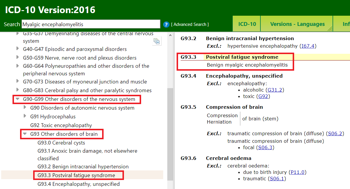
Where can one find ICD 10 diagnosis codes?
Search the full ICD-10 catalog by:
- Code
- Code Descriptions
- Clinical Terms or Synonyms
What are the new ICD 10 codes?
The new codes are for describing the infusion of tixagevimab and cilgavimab monoclonal antibody (code XW023X7), and the infusion of other new technology monoclonal antibody (code XW023Y7).
What are ICD-10 diagnostic codes?
ICD-10-CM Diagnosis Codes
| A00.0 | B99.9 | 1. Certain infectious and parasitic dise ... |
| C00.0 | D49.9 | 2. Neoplasms (C00-D49) |
| D50.0 | D89.9 | 3. Diseases of the blood and blood-formi ... |
| E00.0 | E89.89 | 4. Endocrine, nutritional and metabolic ... |
| F01.50 | F99 | 5. Mental, Behavioral and Neurodevelopme ... |
How many ICD 10 codes are there?
- ICD-10 codes were developed by the World Health Organization (WHO) External file_external .
- ICD-10-CM codes were developed and are maintained by CDC’s National Center for Health Statistics under authorization by the WHO.
- ICD-10-PCS codes External file_external were developed and are maintained by Centers for Medicare and Medicaid Services. ...

What is the geographic atrophy?
Geographic atrophy is a chronic progressive degeneration of the macula and can be seen as part of late-stage age-related macular degeneration (AMD). The condition leads to central scotomas and permanent loss of visual acuity.
Why is geographic atrophy called?
Some patients with age-related macular degeneration (AMD) will develop geographic atrophy (GA), which refers to regions of the retina where cells waste away and die (atrophy). Sometimes these regions of atrophy look like a map to the doctor who is examining the retina, hence the term geographic atrophy.
Is geographic atrophy the same as dry AMD?
Geographic atrophy (GA) is an advanced form of dry age-related macular degeneration (commonly referred to as AMD). AMD is a disease that affects part of the back of the eye called the macula.
What is the prevalence of geographic atrophy?
More than 5 million people worldwide have geographic atrophy, 2 including nearly 1 million people in the U.S. In developed nations, approximately 1 in 29 people over age 75 have geographic atrophy, 3,4,5 which increases to nearly 1 in 4 people over age 90.
What does geographic atrophy look like on OCT?
SD-OCT scans of geographic atrophy, reveals RPE thinning, loss of EPIS and COST lines, depression of the inner retinal layers as the outer layers are loss, and increase visibility of Bruch's membrane and the choroid.
What is nascent Ga?
Nascent GA is a recently described entity, based on specific OCT features that precede manifestations on clinical examination or other imaging modalities and portend the development of drusen-associated GA. From: Atlas of Retinal OCT: Optical Coherence Tomography, 2018.
Is macular atrophy the same as macular degeneration?
Macular atrophy (MA) is a common occurrence during treatment of neovascular age-related macular degeneration (nAMD) with intravitreal anti-vascular endothelial growth factor (VEGF) therapy.
What is dry AMD?
Dry AMD is when parts of the macula get thinner with age and tiny clumps of protein called drusen grow. You slowly lose central vision. There is no way to treat dry AMD yet.
What is atrophic macular degeneration?
Abstract. Atrophic age-related macular degeneration (AMD) is a degenerative disorder of the central retina that greatly increases with age. This complex disease is mainly based on a genetic background but environmental factors are most likely responsible for modulating the individual course of the disorder.
Does geographic atrophy cause total blindness?
Introduction. Geographic atrophy (GA) is considered the late stage of the dry form of age-related macular degeneration (AMD)(1). GA is less common than neovascular AMD and it is responsible for 10-20% of cases of legal blindness in this condition(2,3,4), affecting more than 5 million people wordwilde(5).
How quickly does geographic atrophy progress?
Overall, GA progression rates reported in the literature for total study populations range from 0.53 to 2.6 mm2/year (median, ∼1.78 mm2/year), assessed primarily by color fundus photography or fundus autofluorescence (FAF) imaging.
What is the difference between wet and dry macular degeneration?
The main difference between wet vs dry macular degeneration is simple: dry macular degeneration is the more common type of eye disease and does less damage to your vision while wet macular degeneration can result in serious vision loss.
Coding For Laterality in AMD
When you use the codes for dry AMD (H35.31xx) and wet AMD (H35.32xx), you must use the sixth character to indicate laterality as follows:1 for the...
Coding For Staging in Dry AMD
The codes for dry AMD—H35.31xx—use the seventh character to indicate staging as follows:H35.31x1 for early dry AMD—a combination of multiple small...
Defining Geographic Atrophy
When is the retina considered atrophic? The Academy Preferred Practice Pattern1 defines GA as follows:The phenotype of central geographic atrophy,...
Coding For Geographic Atrophy
The Academy recommends that when coding, you indicate whether the GA involves the center of the fovea: Code H35.31x4 if it does and H35.31x3 if it...
Coding For Staging in Wet AMD
The codes for wet AMD—H35.32xx—use the sixth character to indicate laterality and the seventh character to indicate staging as follows:H35.32x1 for...
When will the ICd 10-CM H35.30 be released?
The 2022 edition of ICD-10-CM H35.30 became effective on October 1, 2021.
What is right macular degeneration?
Right macular degeneration. Clinical Information. A condition in which parts of the eye cells degenerate, resulting in blurred vision and ultimately blindness. A condition in which there is a slow breakdown of cells in the center of the retina (the light-sensitive layers of nerve tissue at the back of the eye).
When will the ICD-10-CM H47.2 be released?
The 2022 edition of ICD-10-CM H47.2 became effective on October 1, 2021.
What is optic disk deficiency?
This condition indicates a deficiency in the number of nerve fibers which arise in the retina and converge to form the optic disk; optic nerve; optic chiasm; and optic tracts.

Popular Posts:
- 1. icd 10 code for male low testosterone
- 2. icd 10 code for intestinal metaplasia of the stomach
- 3. icd 10 code for stenotrophomonas
- 4. icd 10 code for k44.9
- 5. icd 9 code for fibrocystic disease of breast, bilateral?
- 6. icd 10 code for spectacles status
- 7. icd 10 cm code for pelvic cavity hematoma
- 8. icd 10 code for medical diagnosis ptsd
- 9. icd 10 code for cyst of pineal gland
- 10. icd 10 cm code for other long term drug therapy