4.
What is the ICD 10 code for glioblastoma?
Search Page 1/1: glioblastoma. 8 result found: ICD-10-CM Diagnosis Code C71.0 [convert to ICD-9-CM] Malignant neoplasm of cerebrum, except lobes and ventricles. Anaplastic astrocytoma, cerebrum; Astrocytoma of cerebrum; Cancer of the brain, cerebrum, astrocytoma; Cancer of the brain, cerebrum, ependymoma; Cancer of the brain, cerebrum, ...
What is Grade IV glioblastoma?
Glioblastoma (grade IV) Glioblastoma is a highly malignant brain tumor that arises from astrocytes, the supportive cells in the nervous system. Normally, astrocytes are responsible for a variety of roles, including providing nutrients to neurons, maintaining the blood-brain barrier, and modulating neurotransmission...
What is the pathophysiology of glioblastoma?
Glioblastomas often develop in the cerebral hemispheres of the brain, but may occur in almost any area of the brain or spinal cord. They are especially malignant, given that the tumor cells proliferate quickly, and are supported by an extensive network of blood vessels.
What are the genetic signatures of glioblastoma?
Specifically, glioblastomas can have abnormal genetic signatures, including mutations in the IDH1 or IDH2 genes. The presence of these genetic differences can affect prognosis and treatment, and are classified accordingly: What are the treatment options for glioblastoma?
What is the ICD-10 code for glioblastoma?
Malignant neoplasm of brain, unspecified The 2022 edition of ICD-10-CM C71. 9 became effective on October 1, 2021.
What is the ICD-10 code for brain tumor?
ICD-10-CM Code for Malignant neoplasm of brain, unspecified C71. 9.
Is glioma the same as glioblastoma?
A glioma is one of the most common categories of primary brain tumor. Glioblastoma is a type of glioma. Glioma is an umbrella term for cancer of the glial cells that surround nerve endings in the brain.
What is the ICD-10 code for IV?
Encounter for adjustment and management of vascular access device. Z45. 2 is a billable/specific ICD-10-CM code that can be used to indicate a diagnosis for reimbursement purposes. The 2022 edition of ICD-10-CM Z45.
Is glioblastoma malignant?
Glioblastoma is the most common malignant brain and other CNS tumors accounting for 47.7% of all cases. Glioblastoma has an incidence of 3.21 per 100,000 population.
What does GBM stand for in medical terms?
Glioblastoma usually occurs in adults and affects the brain more often than the spinal cord. Also called GBM, glioblastoma multiforme, and grade IV astrocytoma.
What is the difference between Grade 3 & Grade 4 glioma?
Grade 3 brain cancer: The tumor grows quickly, is likely to spread into nearby tissues, and the tumor cells look very different from normal cells. Grade 4 brain cancer: The tumor grows and spreads very quickly, and the tumor cells do not look like normal cells.
How many grades of glioblastoma are there?
Gliomas are classified into four grades (I, II, III, and IV), and the treatment and prognosis depend upon the tumor grade. Astrocytomas, oligodendrogliomas, and glioblastomas are further classified based on whether they have a genetic change in the isocitrate dehydrogenase (IDH) gene.
What causes glioblastoma stage 4?
The causes of glioblastoma are largely unknown. However, it often occurs in people with rare genetic conditions - Turcot syndrome, neurofibromatosis type 1 and Li Fraumeni syndrome - due to mutations in a specific gene that causes many of the characteristic features of glioblastoma.
What is diagnosis code Z51 81?
ICD-10 code Z51. 81 for Encounter for therapeutic drug level monitoring is a medical classification as listed by WHO under the range - Factors influencing health status and contact with health services .
What is the CPT code for IV therapy?
information. According to the American Medical Association (AMA), CPT code 96360 is used to report intravenous (IV) infusions for hydration purposes. The code is used to report the first 31 minutes to 1 hour of hydration therapy.
What is the ICD-10-PCS code for IV hydration?
The objective of this article is to examine the coding of hydration with CPT® codes 96360, Intravenous infusion, hydration; initial, 31 minutes to 1 hour, and 96361, Intravenous infusion, hydration; each additional hour. The purpose of hydration intravenous (IV) infusion is to hydrate.
What is the code for a primary malignant neoplasm?
A primary malignant neoplasm that overlaps two or more contiguous (next to each other) sites should be classified to the subcategory/code .8 ('overlapping lesion'), unless the combination is specifically indexed elsewhere.
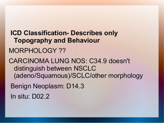
What chapter is neoplasms classified in?
All neoplasms are classified in this chapter, whether they are functionally active or not. An additional code from Chapter 4 may be used, to identify functional activity associated with any neoplasm. Morphology [Histology] Chapter 2 classifies neoplasms primarily by site (topography), with broad groupings for behavior, malignant, in situ, benign, ...
What is a malignant neoplasm?
Malignant neoplasms of ectopic tissue are to be coded to the site mentioned, e.g., ectopic pancreatic malignant neoplasms are coded to pancreas, unspecified ( C25.9 ). A primary or metastatic malignant neoplasm affecting the brain. Cancer of the brain is usually called a brain tumor. There are two main types.
What is oligodendroglioma?
Oligodendroglioma of brain. Primary malignant neoplasm of brain. Primitive neuroectodermal tumor. Secondary malignant neoplasm of spinal cord from neoplasm of brain. Clinical Information. A primary or metastatic malignant neoplasm affecting the brain. Cancer of the brain is usually called a brain tumor.
How do doctors diagnose brain tumors?
doctors diagnose brain tumors by doing a neurologic exam and tests including an mri, ct scan, and biopsy. People with brain tumors have several treatment options. The options are surgery, radiation therapy, and chemotherapy. Many people get a combination of treatments. nih: national cancer institute.
What is the table of neoplasms used for?
The Table of Neoplasms should be used to identify the correct topography code. In a few cases, such as for malignant melanoma and certain neuroendocrine tumors, the morphology (histologic type) is included in the category and codes. Primary malignant neoplasms overlapping site boundaries.
What is the code for a primary malignant neoplasm?
A primary malignant neoplasm that overlaps two or more contiguous (next to each other) sites should be classified to the subcategory/code .8 ('overlapping lesion'), unless the combination is specifically indexed elsewhere.
What is the table of neoplasms used for?
The Table of Neoplasms should be used to identify the correct topography code. In a few cases, such as for malignant melanoma and certain neuroendocrine tumors, the morphology (histologic type) is included in the category and codes. Primary malignant neoplasms overlapping site boundaries.
What is grade IV glioblastoma?
Glioblastoma (grade IV) Glioblastoma is a highly malignant brain tumor that arises from astrocytes, the supportive cells in the nervous system. Normally, astrocytes are responsible for a variety of roles, including providing nutrients to neurons, maintaining the blood-brain barrier, and modulating neurotransmission ...
How common is glioblastoma?
Glioblastomas are the third most common primary brain tumor type, accounting for about 14.9% of primary brain tumors. 1 In 2017, an estimated 12,500 new cases were diagnosed in the United States. 1 Glioblastoma is most common in older adults, but can also occur in children. In children and adolescents, glioblastomas only account for 3% ...
What is the most malignant astrocytoma?
Glioblastomas are the most malignant type of astrocytoma, and also belong to the broader category of gliomas – tumors that arise from glial cells. This is because astrocytes are a type of glial cell. For this reason, glioblastomas may also be called a “grade IV glioma.”. Request an Appointment.
Why is it difficult to remove glioblastoma?
Because glioblastomas tend to spread into neighboring healthy tissue, it can be difficult to fully remove all malignant cells during surgery. This means, that for adult patients, surgery is usually followed by radiation therapy and chemotherapy to target and slow the growth of remaining tumor cells.
What is glioblastoma treated by?
Glioblastoma is a serious condition that will be treated by a multidisciplinary team consisting of neurosurgeons, oncologists, and radiation oncologists. How well a patient with glioblastoma responds to treatment depends on a variety of factors, including the tumor’s size, location, and amount remaining after surgery.
Where do glioblastoma occur?
Glioblastomas often develop in the cerebral hemispheres of the brain, but may occur in almost any area of the brain or spinal cord. They are especially malignant, given that the tumor cells proliferate quickly, and are supported by an extensive network of blood vessels.
Can gamma knife be used on a tumor?
These younger patients usually do not tolerate radiation very well. In some cases, clinicians may be able to use radiotherapies like Gamma Knife that specifically target the tumor site and minimize radiation exposure to the rest of the brain.
Popular Posts:
- 1. icd 10 code for nuclear cataract
- 2. icd 9 code for glioblastoma multiforme
- 3. icd 10 code for severe leukocytosis
- 4. icd 10 code for over lapping toes
- 5. icd-10 code for hiv
- 6. icd code for high index application
- 7. icd 9 code for femoral hematoma
- 8. icd 10 code for wegener granulomatosis
- 9. icd 10 code for increased cholester
- 10. icd 10 code for therapeutic drug monitoring