C71. 1 - Malignant neoplasm of frontal lobe | ICD-10-CM.
What is the ICD 10 code for neoplasm of frontal lobe?
2018/2019 ICD-10-CM Diagnosis Code C71.1. Malignant neoplasm of frontal lobe. 2016 2017 2018 2019 Billable/Specific Code. C71.1 is a billable/specific ICD-10-CM code that can be used to indicate a diagnosis for reimbursement purposes.
What is the ICD 10 code for glioblastoma?
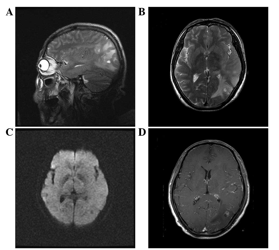
ICD-O: 9445 / 3 - Glioblastoma, IDH mutant ICD-10: C71.1 - Malignant neoplasm of frontal lobe
What is the ICD 10 code for temporal lobe neoplasm?
Malignant neoplasm of temporal lobe. 2016 2017 2018 2019 Billable/Specific Code. C71.2 is a billable/specific ICD-10-CM code that can be used to indicate a diagnosis for reimbursement purposes. The 2018/2019 edition of ICD-10-CM C71.2 became effective on October 1, 2018.
What is the ICD 10 diagnosis group for oligodendroglioma of frontal lobe?
Primary oligodendroglioma of frontal lobe ICD-10-CM C71.1 is grouped within Diagnostic Related Group (s) (MS-DRG v38.0): 054 Nervous system neoplasms with mcc 055 Nervous system neoplasms without mcc
What is the icd10 code for glioblastoma?
9: Malignant neoplasm of brain, unspecified.
What is frontal glioblastoma?
Glioblastoma (GBM), also referred to as a grade IV astrocytoma, is a fast-growing and aggressive brain tumor. It invades the nearby brain tissue, but generally does not spread to distant organs. GBMs can arise in the brain de novo or evolve from lower-grade astrocytoma.
What is the ICD-10-CM code for brain tumor?
ICD-10-CM Code for Malignant neoplasm of brain, unspecified C71. 9.
What is diagnosis code C50 911?
Breast Cancer ICD-10 Code Reference SheetFEMALERightC50.811Malignant neoplasm of overlapping sites, right female breastC50.911Malignant neoplasm of unspecified site, right female breastD05.01Lobular carcinoma in situ, right breast9 more rows
What type of tumors are frontal lobe?
The frontal lobes of the brain are notoriously “silent”: Benign tumors such as meningiomas that compress the frontal lobes from the outside may not produce any symptoms other than progressive change of personality and intellect until they are large.
Is a glioma the same as a glioblastoma?
A glioma is one of the most common categories of primary brain tumor. Glioblastoma is a type of glioma. Glioma is an umbrella term for cancer of the glial cells that surround nerve endings in the brain.
What is the ICD 9 code for brain tumor?
ICD-9 Code 191.9 -Malignant neoplasm of brain unspecified site- Codify by AAPC.
Which of these is a malignant tumor of the brain?
Cancerous (malignant) brain tumors Astrocytoma: These tumors are the most common type of glioma. They form in the star-shaped glial cells called astrocytes. They can form in many parts of your brain, but most commonly occur in your cerebrum. Ependymomas: These tumors often occur near the ventricles in your brain.
What is glioma tumor?
Glioma is a common type of tumor originating in the brain. About 33 percent of all brain tumors are gliomas, which originate in the glial cells that surround and support neurons in the brain, including astrocytes, oligodendrocytes and ependymal cells.
What does c50 912 mean?
912 - Malignant neoplasm of unspecified site of left female breast.
What is primary malignant neoplasm?
A malignant tumor at the original site of growth. [ from NCI]
What is a malignant neoplasm of unspecified site?
A malignant neoplasm (NEE-oh-plaz-um) is another term for a cancerous tumor. The term “neoplasm” refers to an abnormal growth of tissue. The term “malignant” means the tumor is cancerous and is likely to spread (metastasize) beyond its point of origin.
What is the life expectancy of someone with glioblastoma?
The average life expectancy for glioblastoma patients who undergo treatment is 12-15 months and only four months for those who do not receive treatment. Glioblastomas develop from glial cells in the brain and spinal cord.
How serious is a frontal lobe tumor?
Frontal lobe tumors may cause: behavioral and emotional changes; impaired judgment, motivation or inhibition; impaired sense of smell or vision loss; paralysis on one side of the body; reduced mental abilities and memory loss.
How long can you live with a frontal lobe tumor?
The 10-year survival rate is almost 31%. Age is a factor in general survival rates after a cancerous brain or CNS tumor is diagnosed. The 5-year survival rate for people younger than age 15 is about 75%. For people age 15 to 39, the 5-year survival rate nears 72%.
What is the survival rate of glioblastoma?
Glioblastoma Facts & Figures It is estimated that more than 10,000 individuals in the United States will succumb to glioblastoma every year. The five-year survival rate for glioblastoma patients is only 6.8 percent, and the average length of survival for glioblastoma patients is estimated to be only 8 months.
What is the ICd 10 code for glioblastoma?
C71.1 is a billable diagnosis code used to specify a medical diagnosis of malignant neoplasm of frontal lobe. The code C71.1 is valid during the fiscal year 2021 from October 01, 2020 through September 30, 2021 for the submission of HIPAA-covered transactions.#N#The ICD-10-CM code C71.1 might also be used to specify conditions or terms like glioblastoma multiforme, glioblastoma multiforme of brain, glioblastoma multiforme of central nervous system, malignant glioma of cerebrum, malignant neoplasm of frontal lobe , neoplasm of frontal lobe, etc.#N#The following anatomical sites found in the Table of Neoplasms apply to this code given the correct histological behavior: Neoplasm, neoplastic brain NEC frontal lobe or Neoplasm, neoplastic frontal lobe, brain or Neoplasm, neoplastic frontal pole or Neoplasm, neoplastic pole or Neoplasm, neoplastic pole frontal .
What are the different types of glioblastoma?
The following clinical terms are approximate synonyms or lay terms that might be used to identify the correct diagnosis code: 1 Glioblastoma multiforme 2 Glioblastoma multiforme of brain 3 Glioblastoma multiforme of central nervous system 4 Malignant glioma of cerebrum 5 Malignant neoplasm of frontal lobe 6 Neoplasm of frontal lobe 7 Primary glioblastoma multiforme of frontal lobe 8 Primary malignant neoplasm of frontal lobe
How do doctors diagnose brain tumors?
Doctors diagnose brain tumors by doing a neurologic exam and tests including an MRI, CT scan, and biopsy. Treatment options include watchful waiting, surgery, radiation therapy, chemotherapy, and targeted therapy. Targeted therapy uses drugs or other substances that attack cancer cells with less harm to normal cells. Many people get a combination of treatments.
What is a tumor in the brain?
A brain tumor is a growth of abnormal cells in the tissues of the brain. Brain tumors can be benign, with no cancer cells, or malignant, with cancer cells that grow quickly. Some are primary brain tumors, which start in the brain. Others are metastatic, and they start somewhere else in the body and move to the brain.
When was the ICd 10 code implemented?
FY 2016 - New Code, effective from 10/1/2015 through 9/30/2016 (First year ICD-10-CM implemented into the HIPAA code set)
What is a malignant neoplasm?
Malignant neoplasms of ectopic tissue are to be coded to the site mentioned, e.g., ectopic pancreatic malignant neoplasms are coded to pancreas, unspecified ( C25.9 ). A primary or metastatic malignant neoplasm affecting the brain. Cancer of the brain is usually called a brain tumor. There are two main types.
What is oligodendroglioma?
Oligodendroglioma of brain. Primary malignant neoplasm of brain. Primitive neuroectodermal tumor. Secondary malignant neoplasm of spinal cord from neoplasm of brain. Clinical Information. A primary or metastatic malignant neoplasm affecting the brain. Cancer of the brain is usually called a brain tumor.
What is the code for a primary malignant neoplasm?
A primary malignant neoplasm that overlaps two or more contiguous (next to each other) sites should be classified to the subcategory/code .8 ('overlapping lesion'), unless the combination is specifically indexed elsewhere.
How do doctors diagnose brain tumors?
doctors diagnose brain tumors by doing a neurologic exam and tests including an mri, ct scan, and biopsy. People with brain tumors have several treatment options. The options are surgery, radiation therapy, and chemotherapy. Many people get a combination of treatments. nih: national cancer institute.
What is the table of neoplasms used for?
The Table of Neoplasms should be used to identify the correct topography code. In a few cases, such as for malignant melanoma and certain neuroendocrine tumors, the morphology (histologic type) is included in the category and codes. Primary malignant neoplasms overlapping site boundaries.
What chapter is neoplasms classified in?
All neoplasms are classified in this chapter, whether they are functionally active or not. An additional code from Chapter 4 may be used, to identify functional activity associated with any neoplasm. Morphology [Histology] Chapter 2 classifies neoplasms primarily by site (topography), with broad groupings for behavior, malignant, in situ, benign, ...
Where does a brain tumor start?
A primary brain tumor starts in the brain. A metastatic brain tumor starts somewhere else in the body and moves to the brain. Brain tumors can be benign, with no cancer cells, or malignant, with cancer cells that grow quickly.brain tumors can cause many symptoms. Some of the most common are.
The ICD code C71 is used to code Atypical teratoid rhabdoid tumor
Atypical teratoid rhabdoid tumor (AT/RT) is a rare tumor usually diagnosed in childhood. Although usually a brain tumor, AT/RT can occur anywhere in the central nervous system (CNS) including the spinal cord. About 60% will be in the posterior cranial fossa (particularly the cerebellum).
ICD-10-CM Neoplasms Index References for 'C71.1 - Malignant neoplasm of frontal lobe'
The ICD-10-CM Neoplasms Index links the below-listed medical terms to the ICD code C71.1. Click on any term below to browse the neoplasms index.
Equivalent ICD-9 Code GENERAL EQUIVALENCE MAPPINGS (GEM)
This is the official exact match mapping between ICD9 and ICD10, as provided by the General Equivalency mapping crosswalk. This means that in all cases where the ICD9 code 191.1 was previously used, C71.1 is the appropriate modern ICD10 code.
What is the code for a primary malignant neoplasm?
A primary malignant neoplasm that overlaps two or more contiguous (next to each other) sites should be classified to the subcategory/code .8 ('overlapping lesion'), unless the combination is specifically indexed elsewhere.
What chapter is functional activity?
Functional activity. All neoplasms are classified in this chapter, whether they are functionally active or not. An additional code from Chapter 4 may be used, to identify functional activity associated with any neoplasm. Morphology [Histology]
What is the table of neoplasms used for?
The Table of Neoplasms should be used to identify the correct topography code. In a few cases, such as for malignant melanoma and certain neuroendocrine tumors, the morphology (histologic type) is included in the category and codes. Primary malignant neoplasms overlapping site boundaries.
When will the ICD-10 C71.3 be released?
The 2022 edition of ICD-10-CM C71.3 became effective on October 1, 2021.
What is the code for a primary malignant neoplasm?
A primary malignant neoplasm that overlaps two or more contiguous (next to each other) sites should be classified to the subcategory/code .8 ('overlapping lesion'), unless the combination is specifically indexed elsewhere.
What chapter is functional activity?
Functional activity. All neoplasms are classified in this chapter, whether they are functionally active or not. An additional code from Chapter 4 may be used, to identify functional activity associated with any neoplasm. Morphology [Histology]
What is the table of neoplasms used for?
The Table of Neoplasms should be used to identify the correct topography code. In a few cases, such as for malignant melanoma and certain neuroendocrine tumors, the morphology (histologic type) is included in the category and codes. Primary malignant neoplasms overlapping site boundaries.
When will the ICd 10 C71.2 be released?
The 2022 edition of ICD-10-CM C71.2 became effective on October 1, 2021.
What is secondary glioblastoma?
Secondary glioblastoma: IDH mutant glioblastomas constitute the majority of glioblastomas arising from either a diffuse astrocytoma (WHO grade II) or anaplastic astrocytoma (WHO grade III); term not currently in use in pathology ( Clin Cancer Res 2009;15:6002, Science 2008;321:1807 )
Is myxoid morphology variable?
Myxoid background and microcyst formation may be present. Cellular morphology is variable, even within a single tumor. Commonly there is a mix of cells with elongated nuclei and fine fibrillar processes, cells with eccentric nuclei and glassy eosinophilic cytoplasm (gemistocytes), larger pleomorphic cells and small cells with scant cytoplasm.
Can IDH mutant glioblastoma be distinguished from IDH wildtype glioblastom?
IDH mutant glioblastoma cannot be distinguished from IDH wildtype glioblastoma on histology alone.

Popular Posts:
- 1. icd 10 code for diffusion rash
- 2. icd 10 code for breast cyst
- 3. 2019 icd 10 code for ms flaire up
- 4. icd 10 code for dm retinopathy with nonproliferative macular edema
- 5. icd 10 code for hemoperitoneum traumatic
- 6. icd 10 code for sesamoiditis foot
- 7. icd 10 code for strain of left quadriceps muscle fascia and tendon
- 8. icd-10 code for gist
- 9. icd 10 code for strain of thoracic back pain
- 10. 2017 icd 10 code for thin sclerosis periphery