L85. 8 - Other specified epidermal thickening | ICD-10-CM.
What is the ICD 10 Index for keratoacanthoma?
Term: "Keratoacanthoma - ICD-10-CM Index to Diseases and Injuries" "Keratoacanthoma" References in the ICD-10-CM Index to Diseases and Injuries References in the ICD-10-CM Index to Diseases and Injuries applicable to the clinical term "keratoacanthoma" Keratoacanthoma - L85.8 Other specified epidermal thickening
What is the ICD 10 code for keratosis?
Diagnosis Index entries containing back-references to L85.8: Cornu cutaneum L85.8 Dyskeratosis L85.8 Horn cutaneous L85.8 Keratoacanthoma L85.8 Keratosis L57.0 ICD-10-CM Diagnosis Code L57.0 Lichen L28.0 ICD-10-CM Diagnosis Code L28.0 Thickening epidermal L85.9 ICD-10-CM Diagnosis Code L85.9
What is a keratoacanthoma 20 skin?
Keratoacanthoma (20) skin [ICD-10 L85.8] A keratoacanthoma is a skin tumor caused by overexposure to the sun in older adults. The tumor grows quickly, but unlike cancer, it usually does not spread to other parts of the body by way of the blood or lymphatic vessels or membranous surfaces.
Is keratoacanthoma an unreliable histological diagnosis?
The question has been raised as to whether keratoacanthoma is an unreliable histological diagnosis or these tumors have a latent, albeit rare, malignant potential. To date, just a handful of metastasizing keratoacanthomas have been reported.

What is the ICD-10 code for atypical squamous proliferation of skin?
Squamous cell carcinoma of skin, unspecified C44. 92 is a billable/specific ICD-10-CM code that can be used to indicate a diagnosis for reimbursement purposes. The 2022 edition of ICD-10-CM C44. 92 became effective on October 1, 2021.
What is other specified epidermal thickening?
8 for Other specified epidermal thickening is a medical classification as listed by WHO under the range - Diseases of the skin and subcutaneous tissue .
What is the ICD-10 code for skin lesion?
ICD-10-CM Code for Disorder of the skin and subcutaneous tissue, unspecified L98. 9.
What is the ICD-10 code for suspicious lesion?
ICD-10-CM Diagnosis Code B08 B08.
What is the ICD-10 code for sebaceous hyperplasia?
L72. 3 is a billable/specific ICD-10-CM code that can be used to indicate a diagnosis for reimbursement purposes. The 2022 edition of ICD-10-CM L72. 3 became effective on October 1, 2021.
What is the ICD-10 code for actinic keratosis?
ICD-10 code L57. 0 for Actinic keratosis is a medical classification as listed by WHO under the range - Diseases of the skin and subcutaneous tissue .
What's the ICD-10-CM code for sebaceous cyst?
ICD-10 code L72. 3 for Sebaceous cyst is a medical classification as listed by WHO under the range - Diseases of the skin and subcutaneous tissue .
What is the ICD-10 L98 9?
ICD-10 code: L98. 9 Disorder of skin and subcutaneous tissue, unspecified.
What is the ICD-10 code for facial lesion?
Disorder of the skin and subcutaneous tissue, unspecified The 2022 edition of ICD-10-CM L98. 9 became effective on October 1, 2021. This is the American ICD-10-CM version of L98.
What is a ICD code d48 5?
5: Neoplasm of uncertain or unknown behaviour: Skin.
What is the ICD-10 code for skin nodule?
2022 ICD-10-CM Diagnosis Code R22: Localized swelling, mass and lump of skin and subcutaneous tissue.
What is the ICD-10 code for skin infection?
ICD-10 Code for Local infection of the skin and subcutaneous tissue, unspecified- L08. 9- Codify by AAPC.
What is KA on the skin?
The defining characteristic of KA is that it is dome-shaped, symmetrical, surrounded by a smooth wall of inflamed skin, and capped with keratin scales and debris. It grows rapidly, reaching a large size within days or weeks, and if untreated for months will almost always starve itself of nourishment, necrose (die), slough, and heal with scarring. KA is commonly found on sun-exposed skin, often face, forearms and hands.
Can KA be coded as cancer?
You can only code it as cancer if the documentation states its malignancy. "Keratoacanthoma (KA) is a common low-grade (unlikely to metastasize or invade) skin tumour that is believed to originate from the neck of the hair follicle.
Can you code L85.8?
If this were an excision then you will need to wait for a path report before coding. If it were a shave removal or a biopsy then you may code without the path but you will code the L85.8 as the diagnosis. if you have a path report, I would need to know what it states exactly to advise further.
Is keratoacanthoma a malignancy?
Under the microscope, keratoacanthoma very closely resembles squamous cell carcinoma. In order to differentiate between the two, almost the entire structure needs to be removed and examined. While some pathologists classify KA as a distinct entity and not a malignancy, about 6% of clinical and histological keratoacanthomas do progress to invasive and aggressive squamous cell cancers; some pathologists may label KA as "well-differentiated squamous cell carcinoma, keratoacanthoma variant", and prompt definitive surgery may be recommended"
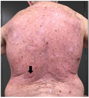
What is the ICd 10 code for keratoacanthoma?
Keratoacanthoma (19) back [ICD-10 L85.8] In A Nutshell. A keratoacanthoma is a skin tumor caused by overexposure to the sun in older adults. The differential diagnosis is squamous cell carcinoma and is always treated the same way by a doctor. The tumor grows quickly, but unlike cancer, it usually does not spread to other parts of the body by way ...
What causes keratoacanthoma?
Trauma, ultraviolet light, chemical carcinogens, human papillomavirus (HPV), genetic factors, and immunocompromised status may trigger keratoacanthoma. It is rare in people younger than 20 years old, and the risk increases significantly after the age of 64.
What are the similarities between keratoacanthoma and squamous cell carcinoma?
Keratoacanthoma and squamous cell carcinoma have similar features, such as actinic damage.
How long does it take for a keratin lesion to stop growing?
It stops growing after 6-8 weeks and remains unchanged for 2-6 weeks. If the core of brown keratin (the material of which hair and the outermost layer of skin is made) comes out, a crate remains. This makes the lesion look like a “mini-volcano.”. The self-healing process then begins.
Where does keratoacanthoma appear?
Symptoms. Keratoacanthoma usually appears on the face, forearms, and hands. It often occurs at site of excessive sun exposure or previous injury or trauma. The lesion is dome-shaped and scaly, while the surrounding skin is smooth but inflamed.
Does keratoacanthoma spread to other parts of the body?
The tumor grows quickly, but unlike cancer, it usually does not spread to other parts of the body by way of the blood or lymphatic vessels or membranous surfaces. Trauma, ultraviolet light, chemical carcinogens, human papillomavirus (HPV), genetic factors, and immunocompromised status may trigger keratoacanthoma.
Does Squamous Cell Carcinoma Go Away On Its Own
They may go away on their own and come back. You should call your doctor if you notice a change in the color, texture, or appearance of your skin or if you have a sore that does not heal or bleeds. Your doctor can diagnose squamous cell carcinoma by examining the growth and performing a biopsy of the suspected area.
What Is The Prognosis Of Keratoacanthoma
The prognosis for keratoacanthoma is excellent following excisional surgery. Recurrent tumors may require more aggressive therapy. Follow patients with a history of keratoacanthoma for development of new primary skin cancers . It has been reclassified as SCC-KA type to reflect the difficulty in histologic differentiation.
Waiting For The Biopsy Result
Wait. And wait. Waiting has been a big part of my skin cancer story. As I waited my tumor grew. Protruding off my forehead was an angry red volcano with a yellow crater in the center. It was still smaller than a dime, but much bigger than the original red bump. I was nervous and worried.
Optimal Therapeutic Approach For This Disease
Management depends on the type of keratoacanthoma, the location, and the number of lesions.
What Are The Symptoms Of Keratoacanthoma
The symptoms of KA are visual and lasts two to three months. The look is often compared to a small volcano.
What Is The Evidence
Nofal, A, Assaf, M, Ghonemy, S, Nofal, E, Yosef, A. âGeneralized eruptive keratoacanthoma: a diagnostic and therapeutic challengeâ. Int J Dermatol. vol. 29. 2014 Jul.
Is Keratoacanthoma Serious
Its a non-melanoma skin cancer that rarely metastasizes, meaning it wont spread to other areas of the body. But it can still be dangerous and should be treated by a doctor. Many people with one KA lesion may develop more throughout their lifetime. But several rare conditions can cause multiple KAs to appear at once.
Is there a good answer to the keratoacanthoma issue?
Take a look at the "similar threads" links just below this. There's no good answer to the keratoacanthoma issue, but those threads will give you a look at previous discussions about it.
Is keratoacanthomas a benign lesion?
I have a coding question, I am hoping you can assist. My physician's experience has been that a keratoacanthoma is a variant of a squamous cell carcinoma (i.e., malignant), so excision of keratoacanthomas would be coded as an excision of a malignant lesion, subtyped by location and size.#N#ICD10 officially lists "keratoacanthoma" as a BENIGN "thickening of the skin", thus medicare (and insurers) would code as a benign excision. Here is a link to some discussion of this topic through AAPC: https://www.aapc.com/memberarea/foru...hickening.html.#N#The pathologist we use (a certified dematopathologist) always reports on whether the margins of a keratoacanthoma are clear or not, which he does only with malignant lesions, not benign lesions. Coders at our office are telling us this is a benign excision.#N#What do you think? Do you think it would be as simple as having the pathologist report it as "keratoacanthoma variant of squamous cell carcinoma" on the report, to show in case of audit? Thank you for your feedback!
Popular Posts:
- 1. icd 9 code for vertebral artery stenosis
- 2. icd 10 code for szr disorders
- 3. icd 10 cm code for allergy to animal
- 4. icd-10 code for pulmonary veing ostial stenosis
- 5. icd 10 code for chronic shin wound
- 6. 2016 icd 10 code for straddle injury
- 7. icd 10 code for subconjunctival hemorrhage of left eye
- 8. icd-10 code for schizophrenia
- 9. icd 10 code for lymptoming
- 10. icd 10 code for abscess of right pinky