ICD-10 code H35. 419 for Lattice degeneration of retina, unspecified eye is a medical classification as listed by WHO under the range - Diseases of the eye and adnexa .
What is the ICD 10 code for bilateral retinal lattice degeneration?
Bilateral retinal lattice degeneration Retinal lattice degeneration, both eyes ICD-10-CM H35.413 is grouped within Diagnostic Related Group (s) (MS-DRG v38.0): 124 Other disorders of the eye with mcc
What is the ICD 10 code for lattice degeneration?
Lattice degeneration of retina, unspecified eye. H35.419 is a billable/specific ICD-10-CM code that can be used to indicate a diagnosis for reimbursement purposes. The 2019 edition of ICD-10-CM H35.419 became effective on October 1, 2018. This is the American ICD-10-CM version of H35.419 - other international versions of ICD-10 H35.419 may differ.
What is the ICD 10 code for hereditary retinal degeneration?
hereditary retinal degeneration (dystrophy) ( ICD-10-CM Diagnosis Code H35.5. Hereditary retinal dystrophy 2016 2017 2018 2019 2020 Non-Billable/Non-Specific Code. Type 1 Excludes dystrophies primarily involving Bruch's membrane (H31.1-) H35.5-)
What is the ICD 10 code for retinal detachment without detachment?
peripheral retinal degeneration with retinal break ( ICD-10-CM Diagnosis Code H33.3. Retinal breaks without detachment 2016 2017 2018 2019 2020 Non-Billable/Non-Specific Code. Type 1 Excludes chorioretinal scars after surgery for detachment (H59.81-) peripheral retinal degeneration without break (H35.4-) H33.3-)
What is lattice degeneration of retina?
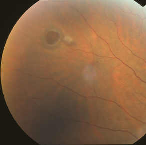
Lattice degeneration is a common peripheral retinal degeneration that is characterized by localized retinal thinning, overlying vitreous liquefaction, and marginal vitreoretinal adhesion. The condition is associated with atrophic retinal holes, retinal tears, and retinal detachments.
Is lattice degeneration a disease?
Lattice degeneration is a condition that affects the retina, which is the tissue at the back of the eye essential for clear and proper vision. Lattice degeneration affects the peripheral portions of the retina, resulting in the tissue developing a lattice pattern.
What causes lattice degeneration of the eye?
Ehlers-Danlos: A genetic connective tissue disease where joints are hyper-extendable and skin is abnormally elastic. This disease is associated with an increased risk of lattice degeneration and retinal detachment. Fundus: The back of the eye where the retina, macula, vitreous, choroid, and optic nerve are located.
How is lattice degeneration diagnosed?
Lattice degeneration itself does not cause symptoms, so the only way to diagnose the condition is with a dilated fundus examination by an eye care provider. A dilated fundus examination is done by administering dilating eye drops in your eyes to expand the pupil so that the retina can be carefully evaluated.
What is lattice in ophthalmology?
Lattice degeneration is a thinning of the retina that happens over time. About 10 percent of people (1 in 10) have lattice degeneration. You need a healthy retina to see clearly, but most with this condition never have any symptoms or a loss in vision. Rarely, lattice degeneration can lead to retinal detachment.
What is the treatment for lattice degeneration?
Lattice degeneration is typically treated with laser to strengthen the retina in areas where it is weak. Side effects are reasonably uncommon, but the risk of side effects increases with the amount of lattice and treatment required. Possible side effects include an increase in pupil size in the treated eye.
Can lattice degeneration cause blindness?
What are symptoms of lattice degeneration? Lattice degeneration does not have any symptoms. But because the retina is thinner with lattice degeneration, it may tear, break, or get holes easier. This can lead to retinal detachment, which can cause blindness without treatment.
Can you have cataract surgery with lattice degeneration?
When a patient with lattice degeneration is considered for cataract surgery, one management option includes prophylactic treatment. Given the low incidence of retinal detachment, it is unnecessary to treat all patients with lattice degeneration prophylactically.
At what age does lattice degeneration start?
Lattice patients with low to moderate degrees of myopia tend to develop detachments between 40 and 60 years of age caused by premature posterior vitreous separation and tractional tears.
Popular Posts:
- 1. icd 10 code for refusal of treatment
- 2. icd 10 code for osteophyte right hand
- 3. icd 10 cm code for cataract due to type 2 diabetes mellitus
- 4. icd 10 code for injury of left toe
- 5. icd 10 code for malpositioned dental appliance
- 6. icd 10 code for focal nodular hyperplasia of liver
- 7. icd 10 code for history of gastric ulcer
- 8. icd 10 code for disc extrusion lumbar
- 9. what is the icd 10 code for staphylococcal pharyngitis
- 10. icd-10-cm code for delivery of twins by cesarean section