Unspecified lump in the left breast, upper outer quadrant
The 2022 edition of ICD-10-CM N63. 21 became effective on October 1, 2021.What is the ICD 10 code for lump in left breast?
Oct 01, 2021 · 2022 ICD-10-CM Diagnosis Code N63.21 Unspecified lump in the left breast, upper outer quadrant 2018 - New Code 2019 2020 2021 2022 Billable/Specific Code N63.21 is a billable/specific ICD-10-CM code that can be used to indicate a diagnosis for reimbursement purposes. The 2022 edition of ICD-10-CM N63.21 became effective on October 1, 2021.
What is the ICD 10 code for breast cancer?
Code the specific quadrant for multifocal tumors all within one quadrant • Do not code C509 (Breast, NOS) in this situation Code the primary site to C508 when . O'Clock Positions and Codes Quadrants of Breasts 2 11 12 1 1 10 9 8 7 7 6 5 4 3 2 11 12 10 6 5 3 RIGHT BREAST LEF T BREAST UOQ UIQ UIQ UOQ LOQ LIQ LIQ LOQ
What is the ICD 10 code for mass and lump?
Oct 01, 2021 · Unspecified lump in the left breast, unspecified quadrant 2018 - New Code 2019 2020 2021 2022 Billable/Specific Code N63.20 is a billable/specific ICD-10-CM code that can be used to indicate a diagnosis for reimbursement purposes. The 2022 edition of ICD-10-CM N63.20 became effective on October 1, 2021.
What is the ICD 10 code for lump in upper quadrant?
Quadrants of the Breast. Note: C50.6 is the code for axillary tail or tail of breast. « Previous (Anatomy) Next (Regional Lymph Nodes) ».
What is the ICD-10 code for Mass in left breast?
Unspecified lump in the left breast, overlapping quadrants N63. 25 is a billable/specific ICD-10-CM code that can be used to indicate a diagnosis for reimbursement purposes.
What is the ICD-10 code for breast mass?
N63. 0 - Unspecified lump in unspecified breast. ICD-10-CM.
What quadrant is 12 o'clock on the breast?
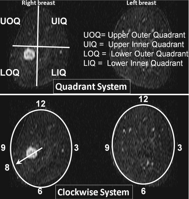
In the left breast the upper outer quadrant is between 12 and 3 o'clock.
What quadrant is 9 o'clock in the breast?
1 Central portion of breast. lower inner quadrant is between 3 and 6 o'clock; the lower outer quadrant is between 6 and 9 o'clock; and the upper outer quadrant is between 9 and 12 o'clock.
What is a mass on the breast?
A breast lump is a mass that develops in the breast. Breast lumps vary in size and texture and may cause pain. Some are not found until a physical or imaging exam. Most breast lumps are benign (non-cancerous).
What happens when a mass is found in the breast?
A lump or mass in the breast is the most common symptom of breast cancer. Lumps are often hard and painless, although some are painful. However, not all lumps are cancer. Benign breast conditions (like cysts) that can also cause lumps.Sep 22, 2020
What does suspicious mass in breast mean?
A mass might be seen with or without calcifications. Masses can be many things, including cysts (non-cancerous, fluid-filled sacs) and non-cancerous solid tumors (such as fibroadenomas), but they may also be a sign of cancer. Cysts are fluid-filled sacs.Jan 14, 2022
What is 12 o'clock position?
LL2218-7Clock positionActive A clock position is the relative direction of an object described using the analogy of a 12-hour clock. For example, 12 o'clock means ahead or above, 3 o'clock means to the right, 6 o'clock means behind or below, and 9 o'clock means to the left.
What quadrant has most breast cancers?
Most breast cancers develop in the upper outer quadrant of the breast, closest to the armpit. This is because this area has a lot of glandular tissue.
Where are cancerous lumps found in the breast?
Commonly developing from the mammary glands or ducts, such malignant lumps generally (about 50 percent) appear in the upper, outer quadrant of the breast, extending into the armpit, where tissue is thicker than elsewhere.
What are the 4 quadrants of the breast?
Classification of Breast Quadrants A single breast can be divided into four quadrants: UO, upper inner (UI), lower outer (LO), and lower inner (LI) by two perpendicular planes intersected at the nipple.
Where are most breast tumors located?
About half of cancerous breast lumps appear in the upper, outer quadrant of the breast, extending into the armpit. About 18 percent of breast cancer tumors show up in the nipple area. Around 11 percent are found in the lower quadrant, and 6 percent are located in the lower, inner quadrant.Apr 14, 2021
What chapter is functional activity?
Functional activity. All neoplasms are classified in this chapter, whether they are functionally active or not. An additional code from Chapter 4 may be used, to identify functional activity associated with any neoplasm. Morphology [Histology]
What is the code for a primary malignant neoplasm?
A primary malignant neoplasm that overlaps two or more contiguous (next to each other) sites should be classified to the subcategory/code .8 ('overlapping lesion'), unless the combination is specifically indexed elsewhere.
What are the sections of a radiology report?
The reports of your radiology exams usually contain three sections: 1 Exam description and history – the type of exam, day it was performed, the reason it was performed and any important patient information 2 Findings – a detailed description of the important findings on the exam including size, shape, location and changes 3 Impression – a summary of the findings, what they mean and what to do about them Radiologists use standard terms in reports to describe the appearance of important findings.
Why do breasts change size?
A number of conditions other than breast cancer can cause breasts to change in size or feel. Breast tissue changes naturally during pregnancy and a woman’s menstrual cycle. Other possible causes of non-cancerous (benign) breast changes include fibrocystic changes, cysts, fibroadenomas, infection or injury.
What does it mean when you have a lump in your breast?
Lump in the breast or in the underarm. A spontaneous or bloody discharge from the nipple. New retraction or indentation of the nipple. A change in the size or contour of the breast. Any flattening or indentation of the skin over the breast. Redness or pitting of the skin over the breast, like the skin of an orange.
How to determine if you have breast cancer?
Essentially, the histological evaluation is the microscopic analysis of the chemical and cellular properties associated with a suspicious breast tumor.
Is breast cancer more common in women?
Breast Cancer is one of the most common and well-known cancers diagnosed in the United States. It can occur in both women and men, but is substantially more common in women.
What is an initial biopsy?
An initial biopsy sampling and analysis could be considered as an extension of the breast cancer screening process, where breast cancer is either confirmed positive or confirmed negative. Once breast cancer is confirmed by the pathologist, the breast cancer staging process begins.
Is breast cancer a lump?
When the disease is discovered early, there are more treatment options and a better chance for a cure. Most painful breast lumps are not cancerous. Any discrete breast lump whether painful or not should be evaluated because breast cancer often presents as a lump or thi ckening.

Popular Posts:
- 1. icd 10 code for encounter for removal of surgical dressing change
- 2. icd 10 code for redness throat
- 3. icd 10 code for renal pain
- 4. icd 10 code for hcg testing
- 5. icd 10 code for abrasion left great toe
- 6. icd 10 code for insulin dependent type 2 diabetes
- 7. icd 10 code for hx of hip fracture
- 8. icd-10 code for blindness
- 9. icd 10 code for staph
- 10. icd 10 code for cervical disc disease unspecified