Displaced fracture of body of hamate [unciform] bone, left wrist, initial encounter for closed fracture. S62. 142A is a billable/specific ICD-10-CM code that can be used to indicate a diagnosis for reimbursement purposes.
Full
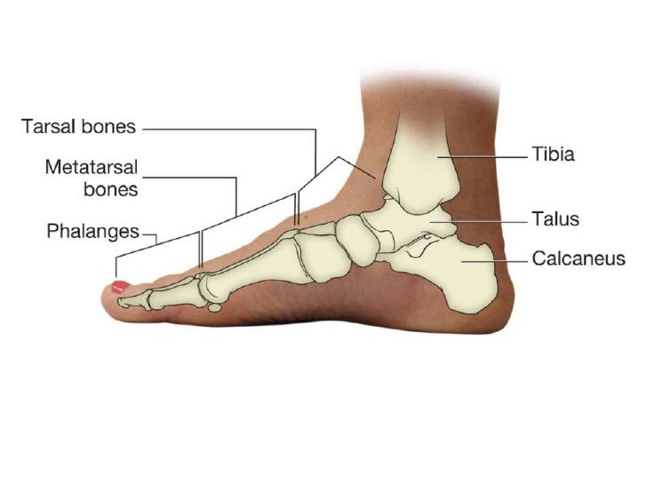
AnswerWhat is a hamate bone?
Hamate bone (left hand) animation. The hamate bone is one of eight carpal bones that forms part of the wrist joint. The word hamate is derived from the Latin word hamulus which means “a little hook”. It is a wedge-shaped bone with a hook-like process that can be found in the medial side of the wrist.
What is hook hamate fracture?
Hook of hamate fracture is a fracture of a hook shaped protrusion on the hamate bone, which is one of the small carpal bones in the wrist. It may occur when swinging a golf club or tennis racket against an immovable object.
How is a hamate fracture caused?
The body of the hamate fracture is a consequence of a direct blow over the hypothenar eminence or a considerably strong dorsopalmar compression. [3][14] A body fracture may also accompany high energy trauma resulting in wrist fracture dislocations. [15] Body fractures can lead to axial carpal instability.
What happens if you break your hamate bone?
The most common symptom of a fracture of the hook of the hamate is pain. Swelling, bruising, and weakness of grip are also common. The pain may be vague and difficult to reproduce, but should be found when an examiner presses directly on the hook of the hamate bone.
What is hamate surgery?
The typical surgical procedure for Hook of Hamate fractures and nonunions involves removing the fractured or non-united “hook”. Although rarely, standard fracture fixation with screws may be performed to fix the fracture.
What goes through the hook of hamate?
The pisiform-hamate ligament, flexor retinaculum (also known as the transverse carpal ligament), flexor carpi ulnaris tendon, opponens digiti minimi tendon, and flexor digiti minimi tendon all attach to the hook.
How is a hamate fracture diagnosed?
Imaging can assist with diagnosis of these injuries. The overlap of the hook of the hamate on the body can lead to difficulty picking up these fractures on standard hand series radiographs. Often a carpal tunnel view or supinated oblique view can better identify the fracture.
What is the function of the hamate?
The main function of this bone is to connect the distal row of carpal bones with the fourth and fifth metacarpal bones. The hamate bone serves as the attachment point for a number of muscles and ligaments of the hand and forearm. In addition, it participates in the formation of the carpal tunnel and Guyon's canal.
What is Bennett's fracture?
The Bennett fracture is the most common fracture involving the base of the thumb. This fracture refers to an intraarticular fracture that separates the palmar ulnar aspect of the first metacarpal base from the remaining first metacarpal.
How is hamate injury treated?
Conservative treatment requires immobilization with casting for 6 weeks, followed by an additional 4-6 weeks of physical therapy. If the injury is treated surgically with hook excision, the patient can start physical therapy immediately, without limitations, and can return to full activity within 6-8 weeks.
How long does a broken hamate take to heal?
Typically, if treated conservatively, simple fractures of the hamate are unified within 6-8 weeks of injury. Patient participation in full-contact sports, such as football, usually requires bracing or protection for the wrist until full musculature and flexibility have returned.
How do baseball players break their hamate bone?
The hamate bone is one of eight carpal bones in the wrist, and is located on the outside of the pinky finger. A fracture to the bone frequently occurs in baseball and golf or in racquet sports due to the way the bat (club or racquet handle) makes contact and exerts pressure on the bone.
What is the ICd 10 code for hamate fracture?
Fracture of body of hamate [unciform] bone 1 S62.14 should not be used for reimbursement purposes as there are multiple codes below it that contain a greater level of detail. 2 The 2021 edition of ICD-10-CM S62.14 became effective on October 1, 2020. 3 This is the American ICD-10-CM version of S62.14 - other international versions of ICD-10 S62.14 may differ.
What is the secondary code for Chapter 20?
Use secondary code (s) from Chapter 20, External causes of morbidity, to indicate cause of injury. Codes within the T section that include the external cause do not require an additional external cause code. Type 1 Excludes.
Popular Posts:
- 1. icd 10 code for history intracerebral hemorrhage
- 2. icd 10 code for gas embolism
- 3. icd 10 code for elevated calprotectin
- 4. icd 10 code for l5 fracture
- 5. icd 10 code for left zygomaticomaxillary fracture
- 6. 2017 icd 10 code for prostate cancer metastatic to bone
- 7. icd 10 dx code for bilateral ear pain
- 8. 2015 icd 10 code for c abg times 6
- 9. icd 10 code for obstructive sleep apnea syndrome
- 10. icd 10 code for loss of hearing right ear