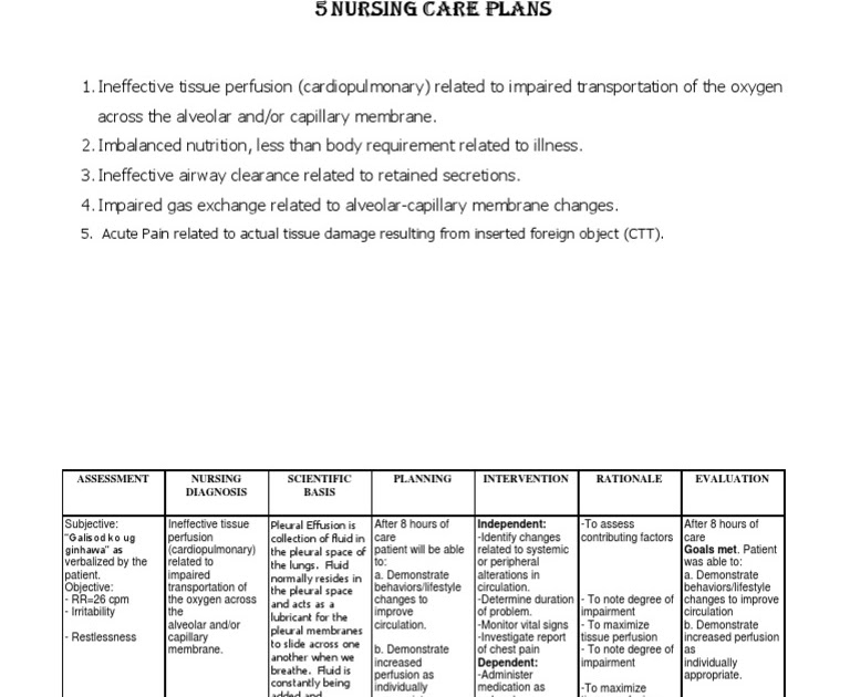
J91. 8 - Pleural effusion in other conditions classified elsewhere | ICD-10-CM.
What increases my risk for pleural effusion?
The following diseases may cause pleural effusion:
- Heart failure
- Bacterial pneumonia
- lung cancer and other tumours with lung metastases
- Pulmonary embolism
- Radiation therapy to the chest
- Nephrotic syndrome
- Hypothyroidism
- Ovarian tumours
- Tuberculosis
- Connective tissue disease (for example, rheumatoid arthritis, lupus)
What are the signs of pleural effusion?
Signs and symptoms of a pleural effusion include chest pain, shortness of breath or difficulty breathing, asymmetrical expansion of the chest during breathing, and a dry or productive (producing sputum) cough. Other associated symptoms can include pleurisy, which is pain in the chest that occur during breathing.
What is the diagnosis code for pleural effusion?
- chylous (pleural) effusion ( ICD-10-CM Diagnosis Code J94.0. Chylous effusion.
- malignant pleural effusion ( ICD-10-CM Diagnosis Code J91.0. Malignant pleural effusion.
- pleurisy NOS ( ICD-10-CM Diagnosis Code R09.1. Pleurisy.
- tuberculous pleural effusion ( ICD-10-CM Diagnosis Code A15.6. Tuberculous pleurisy.
What does no evidence of pleural effusion mean?
“No sizeable pleural effusion or pneumothorax identified” makes more sense. Which means neither a pleural effusion or pneumothorax is seen. The use of the word “sizeable” could just be a speaking style or could be suggesting there are some minor limitations to the xrays.
What is the ICD-10-CM code for pleural effusion?
ICD-10 code J90 for Pleural effusion, not elsewhere classified is a medical classification as listed by WHO under the range - Diseases of the respiratory system .
What is left side pleural effusion?
What is pleural effusion? Pleural effusion, sometimes referred to as “water on the lungs,” is the build-up of excess fluid between the layers of the pleura outside the lungs. The pleura are thin membranes that line the lungs and the inside of the chest cavity and act to lubricate and facilitate breathing.
Is pleural effusion right or left?
Pleural effusions in patients with congestive heart failure are typically bilateral. However, a unilateral pleural effusion is more commonly seen on the right side.
What is pleural effusion defined as?
(PLOOR-ul eh-FYOO-zhun) An abnormal collection of fluid between the thin layers of tissue (pleura) lining the lung and the wall of the chest cavity.
What are the two types of pleural effusion?
There are two types of pleural effusion:Transudative pleural effusion is caused by fluid leaking into the pleural space. ... Exudative effusion is caused by blocked blood vessels or lymph vessels, inflammation, infection, lung injury, and tumors.
What is small bilateral pleural effusions?
Bilateral pleural effusion is an abnormal accumulation of fluid in the pleural space -- the space between the lungs and the chest wall, said doctors. Advertisement. By: Lifestyle Desk | New Delhi | June 7, 2021 7:10:05 pm. The disease can be diagnosed through X-ray and CT scan of the chest. ( Photo: Getty/Thinkstock)
Is pleural effusion and pneumonia the same thing?
Pleural effusion is a buildup of fluid in the pleural space. The pleural space is the area between the layers of the tissue lining the lung and the chest cavity. In a person with parapneumonic pleural effusion, the fluid buildup is caused by pneumonia.
What is the difference between pleural effusion and pericardial effusion?
Q: Pericardial effusion vs. pleural effusion - what is the difference? A: Pericardial effusion is the term for a buildup of fluid around the heart. Pleural effusion is the term for a buildup of fluid around the lungs, or, more accurately, in the space between the lungs and the chest cavity.
What is pleural effusion or pneumothorax?
Pleural effusion - excess fluid in the pleural space. Pneumothorax - buildup of air or gas in the pleural space. Hemothorax - buildup of blood in the pleural space.
Is effusion the same as swelling?
Effusion is swelling that happens when fluid leaks out of a vein, artery, lymph vessel, or synovial membrane into the surrounding tissue. This causes the tissue to expand, or swell. When effusion happens in a joint — commonly the knee — excess fluid can pool in a part of the joint called the synovial cavity.
Why does pleural effusion occur?
Pleural effusion occurs when fluid builds up in the space between the lung and the chest wall. This can happen for many different reasons, including pneumonia or complications from heart, liver, or kidney disease. Another reason could be as a side effect from cancer.
What is an effusion in medical terms?
Listen to pronunciation. (eh-FYOO-zhun) An abnormal collection of fluid in hollow spaces or between tissues of the body. For example, a pleural effusion is a collection of fluid between the two layers of membrane covering the lungs.
What is the ICd code for pleural effusion?
J90 is a billable ICD code used to specify a diagnosis of pleural effusion, not elsewhere classified. A 'billable code' is detailed enough to be used to specify a medical diagnosis.
What is the term for a fluid that can impair breathing?
Various kinds of pleural effusion, depending on the nature of the fluid and what caused its entry into the pleural space, are hydrothorax (serous fluid), hemothorax (blood), urinothorax (urine), chylothorax (chyle), or pyothorax (pus). Pneumothorax is the accumulation of air ...
What is inclusion term?
Inclusion Terms are a list of concepts for which a specific code is used. The list of Inclusion Terms is useful for determining the correct code in some cases, but the list is not necessarily exhaustive.
The ICD code J91 is used to code Chylothorax
A chylothorax (or chyle leak) is a type of pleural effusion. It results from lymph formed in the digestive system called chyle accumulating in the pleural cavity due to either disruption or obstruction of the thoracic duct.
Equivalent ICD-9 Code GENERAL EQUIVALENCE MAPPINGS (GEM)
This is the official approximate match mapping between ICD9 and ICD10, as provided by the General Equivalency mapping crosswalk. This means that while there is no exact mapping between this ICD10 code J91.8 and a single ICD9 code, 511.9 is an approximate match for comparison and conversion purposes.
How many new CPT codes were released in January?
In January, new CPT codes were released. There were 248 new CPT codes added, 71 deleted and 75 revised. Most of the surgery section changes were in the musculoskeletal and cardiovascular subsections. These included procedures such as skin grafting, breast biopsies, deep drug delivery systems, tricuspid valve repairs, aortic grafts and repair of iliac artery.
How many ICD-10 codes are there for FY2021?
In this part, the ICD-10-PCS procedure codes are presented. For FY2021 ICD-10-PCS there are 78,115 total codes (FY2020 total was 77,571); 556 new codes (734 new last year in FY2020)…
What is a pseudodoseizure?
Pseudoseizures are a form of non-epileptic seizure. These are difficult to diagnose and oftentimes extremely difficult for the patient to comprehend. The term “pseudoseizures” is an older term that is still used today to describe psychogenic nonepileptic seizures (PNES).
What is the purpose of anticoagulant?
Anticoagulants and antiplatelets are used for the prevention and treatment of blood clots that occur in blood vessels. Oftentimes, anticoagulants and antiplatelets are referred to as “blood thinners,” but they don’t actually thin the blood at all. These drugs slow down the body’s process of making clots.
What is the R40.2- scale?
The coma scale codes (R40.2-) can be used in conjunction with traumatic brain injury codes, acute cerebrovascular disease or sequelae of cerebrovascular disease codes. These codes are primarily for use by trauma registries, but they may be used in any setting where this information is collected. The coma scale may also be used to assess the status of the central nervous system for other non-trauma conditions, such as monitoring patients in the intensive care unit regardless of medical condition.
What is the Z20.828 code?
Assign code Z20.828, “Contact with and (suspected) exposure to other viral communicable diseases” for all patients who are tested for COVID-19 and the results are negative, regardless of symptoms, no symptoms, exposure or not as we are in a pandemic.
What is client S?
“Client S” is a small, not-for-profit, 40 bed micro-hospital in the Southeast. HIA performed a 65-record review this year for Client S and found an opportunity with 15 of them. 9 had an increased reimbursement with a total of $43,228 found.
Popular Posts:
- 1. 2019 icd 10 code for periorbital hematoma
- 2. icd code for vaginal yeast infection
- 3. icd 10 code for tortuous carotid artery
- 4. icd 10 cm code for cyst hepatic
- 5. icd-10 code for subclinical hyperthyroidism
- 6. icd 10 code for 371.2
- 7. icd 10 code for scalp edema
- 8. icd 10 code for pain right big toe
- 9. icd 10 code for lgsil of cervix
- 10. icd 10 code for bandage contact lens