Personal history of urinary calculi
Z87. 442 is a billable/specific ICD-10-CM code that can be used to indicate a diagnosis for reimbursement purposes. The 2022 edition of ICD-10-CM Z87. 442 became effective on October 1, 2021.What is the ICD 10 code for calculus of lower urinary tract?
Calculus of lower urinary tract, unspecified. N21.9 is a billable/specific ICD-10-CM code that can be used to indicate a diagnosis for reimbursement purposes. The 2018/2019 edition of ICD-10-CM N21.9 became effective on October 1, 2018.
What is the ICD 10 calculus of kidney 2019?
Calculus of kidney. The 2019 edition of ICD-10-CM N20.0 became effective on October 1, 2018. This is the American ICD-10-CM version of N20.0 - other international versions of ICD-10 N20.0 may differ.
What is the ICD 10 code for kidney cancer?
Calculus of kidney. 2016 2017 2018 2019 2020 Billable/Specific Code. N20.0 is a billable/specific ICD-10-CM code that can be used to indicate a diagnosis for reimbursement purposes. The 2020 edition of ICD-10-CM N20.0 became effective on October 1, 2019.
What is the ICD 10 code for calcification of the kidney?
Diagnosis Index entries containing back-references to N28.89: Abscess (connective tissue) (embolic) (fistulous) (infective) (metastatic) (multiple) (pernicious) (pyogenic) (septic) L02.91 ICD-10-CM Diagnosis Code L02.91 Adhesions, adhesive (postinfective) K66.0 ICD-10-CM Diagnosis Code K66.0 Calcification kidney N28.89 Calicectasis N28.89
What is the ICD-10 code for ESWL?
98.5198.51 Extracorporeal shockwave lithotripsy [ESWL] of the kidney, ureter and/or bladder.
What is the ICD-10 code for left renal calculi?
0.
What is the ICD-10-CM code for Ureterolithiasis?
1: Calculus of ureter.
What is the code N20 0?
0: Calculus of kidney.
What is the procedure for lithotripsy?
Lithotripsy treats kidney stones by sending focused ultrasonic energy or shock waves directly to the stone first located with fluoroscopy (a type of X-ray “movie”) or ultrasound (high frequency sound waves). The shock waves break a large stone into smaller stones that will pass through the urinary system.
What are the types of lithotripsy?
The two main types of lithotripsy are extracorporeal shock wave lithotripsy (ESWL) and laser lithotripsy. Laser lithotripsy is sometimes known as flexible ureteroscopy and laser lithotripsy (FURSL) because doctors use a tool called a ureteroscope.
What is the diagnosis code for kidney stone?
N20. 0 is a billable/specific ICD-10-CM code that can be used to indicate a diagnosis for reimbursement purposes. The 2022 edition of ICD-10-CM N20.
What is right renal Calculus?
Kidney stones (also called renal calculi, nephrolithiasis or urolithiasis) are hard deposits made of minerals and salts that form inside your kidneys. Diet, excess body weight, some medical conditions, and certain supplements and medications are among the many causes of kidney stones.
What is Ureterolithiasis medical term?
Ureterolithiasis, which literally translates to stones in the ureter, is sometimes referred to improperly as “kidney stones,” which are properly known as nephrolithiasis. Although stones do form within the kidney, they do not typically cause acute pain.
What is the ICD-10 code for history of kidney stones?
ICD-10 code Z87. 442 for Personal history of urinary calculi is a medical classification as listed by WHO under the range - Factors influencing health status and contact with health services .
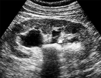
What is the hydronephrosis in kidney?
Hydronephrosis is a condition where one or both kidneys become stretched and swollen as the result of a build-up of urine inside them. It can affect people of any age and is sometimes spotted in unborn babies during routine pregnancy ultrasound scans.
Is nephrolithiasis a disease?
Nephrolithiasis, or kidney stone disease, is a condition in which individuals form calculi (stones) within the renal pelvis and tubular lumens. Stones form from crystals that precipitate (separate) out of the urine.
When do you code hypertensive heart disease?
Heart failure is assumed to be due to hypertension when coded using I11. 0, “Hypertensive heart disease with heart failure.” In ICD-10, the word “with” presumes a causal relationship between the two conditions linked by this term.
What is the ICD-10 for abdominal pain?
ICD-10 code R10. 9 for Unspecified abdominal pain is a medical classification as listed by WHO under the range - Symptoms, signs and abnormal clinical and laboratory findings, not elsewhere classified .
What modifier is used for second ESWL?
Solution: If the second ESWL is for a different stone in the same kidney and done within the postoperative period of the first ESWL Center advises using modifier -79 (Unrelated procedure) to indicate an unrelated procedure. “It’s unrelated to the first surgery” Center says. “Even though it’s the same procedure it’s a different stone.”
What is ESWL 50590?
ESWL represented by 50590 (Lithotripsy extracorporeal shock wave) is one of the most frequently performed procedures in urology practices as well as a popular and effective treatment for renal calculus (kidney stones). Our experts give you the facts for seven tricky ESWL coding scenarios.
What is 50081 ESWL?
After one month the physician realizes the stone was incompletely fragmented by the ESWL and decides to perform 50081 (Percutaneous nephrostolithotomy or pyelostolithotomy with or without dilation endoscopy lithotripsy stenting or basket extraction; over 2 cm). The diagnosis for both procedures is 592.0 (Calculus of kidney).
What modifier do you use for a ureteral stent?
Solution: Medicare views the need for a stent such as a stone obstructing the ureter as a complication so you should append modifier -78 (Return to the operating room for a related procedure during the postoperative period) to 52332 (Cystourethroscopy with insertion of indwelling ureteral stent [e.g. Gibbons or double-J type]) says Karen Delebreau coding specialist with Urological Surgeons in Green Bay Wis.
What modifier is used for a second procedure code?
For commercial and private carriers bill the treatment of this scenario exactly the same appending modifier -78 to the second procedure code. However some private payers might view this as a new problem and therefore require you to append modifier -79 (Unrelated procedure or service by the same physician during the postoperative period) to 52332.
What modifiers are used for kidney stones?
Remember: Use -LT (Left) and -RT (Right) modifiers to indicate which kidney the ESWL targeted Center says. If the left kidney stone is treated first use 50590-LT; for the second ESWL use 50590-RT-58.
Can you charge 50590 for kidney stones?
Solution: Sorry – if there are several stones in one kidney you cannot charge 50590 more than once for that session says Carolyn Zell CPC billing manager for the Urology Team in Austin Texas.
What was instilled in the urethra of a swollen bladder?
The bladder was emptied, and lidocaine jelly was instilled in the urethra. He was extubated and taken to the recovery room in good condition.
What is the diagnosis of bladder cancer?
Postoperative diagnosis: Transitional cell carcinoma in the bladder. (This is the diagnosis to report, since the pre and post-operative diagnoses are the same. The operative note is consistent with a tumor on the posterior bladder wall. Pathology is not back yet, but the stated diagnosis is transitional cell carcinoma in the bladder. In the US, 90% of all bladder cancers are transitional cell in origin. This is sometimes referred to as urothelial carcinoma.)
What is the external ring of the ilioinguinal ligament?
A scalpel was used to make a skin incision following the creases and this was extended down through very generous subcutaneous fat and Scarpa's fascia to expose the external oblique aponeurosis. The external ring was identified as was the ilioinguinal ligament. The ring was opened for a short distance.
What is retrograde pyelogram?
A bilateral retrograde pyelogram was performed, which showed no filling defects or irregularities. (Retrograde radiological imaging (supervision and interpretation) of the kidneys and ureters. Retrograde refers to going against the normal flow. Urine flows down to the bladder and the dye is injected to travel back up towards the kidney.)

Popular Posts:
- 1. icd 9 code for metastatic colon cancer to liver
- 2. icd 10 code for arm discomfort
- 3. icd 10 code for routine colonoscopy
- 4. icd 10 code for painful internal fixation device
- 5. icd-10 code for copd with bronchitis
- 6. icd 10 code for ss recurrent bacteremia
- 7. icd 10 code for pituratary tumor
- 8. icd 10 code for presenile psychosis
- 9. icd 10 cm code for left abd pain
- 10. icd 10 code for leaking gastrostomy tube