ICD-10 code D05. 01 for Lobular carcinoma in situ of right breast is a medical classification as listed by WHO under the range - Neoplasms .
What is the ICD 10 code for breast cancer?
Personal history of malignant neoplasm of breast
- Z85.3 is a billable/specific ICD-10-CM code that can be used to indicate a diagnosis for reimbursement purposes.
- The 2022 edition of ICD-10-CM Z85.3 became effective on October 1, 2021.
- This is the American ICD-10-CM version of Z85.3 - other international versions of ICD-10 Z85.3 may differ.
What is the diagnosis code for breast cancer?
- 174.0, Nipple and areola;
- 174.1, Central portion;
- 174.2, Upper-inner quadrant;
- 174.3, Lower-inner quadrant;
- 174.4, Upper-outer quadrant;
- 174.5, Lower-outer quadrant;
- 174.6, Axillary tail;
Is breast complex cyst sign of cancer?
The term cystic breast lesions is a sonographic diagnosis, which can be categorized as simple, complicated or complex. The complex cysts are deemed as having a low risk of breast cancer. The highly aggressive invasive ductal carcinomas among complex cysts, in particular, are infrequent reported.
Is associated with breast cancer prognosis?
There is a significant increase in the risk of contralateral breast cancers in BRCA mutation carriers with an estimated 10-year risk ranging from 20-40%. The prognosis of BRCA-associated breast cancers appears to be similar to that of sporadic breast cancers based on the current literature. Future data from large prospective cohort studies will ...
What is the ICD 10 code for lobular carcinoma in situ?
D05. 01 is a billable/specific ICD-10-CM code that can be used to indicate a diagnosis for reimbursement purposes. The 2022 edition of ICD-10-CM D05. 01 became effective on October 1, 2021.
What is lobular breast cancer in situ?
Lobular carcinoma in situ (LCIS) is an uncommon condition in which abnormal cells form in the milk glands (lobules) in the breast. LCIS isn't cancer. But being diagnosed with LCIS indicates that you have an increased risk of developing breast cancer.
Is lobular carcinoma in situ bilateral?
LCIS is multicentric in 60-80% of patients 14 and bilateral in 20-60% 11, 15, 16.
Is lobular carcinoma in situ malignant?
These are benign (non-cancerous) conditions, but they both increase your risk of breast cancer. The different types of LCIS are: Classic LCIS: The cells lining the lobules of the breast are smaller and are about the same size.
Why is DCIS considered cancer and LCIS is not?
Overview. Unlike ductal carcinoma in situ or DCIS, LCIS is not considered a precursor to invasive breast cancer so it does not require treatment. If left alone, LCIS does not turn into invasive breast cancer. Rather, LCIS is considered a marker for increased breast cancer risk in either breast, much like family history ...
Is DCIS the same as LCIS?
Lobular carcinoma in situ (LCIS) is a type of in-situ carcinoma of the breast. While DCIS is considered a pre-cancer, it is unclear whether LCIS is definitely a pre-cancer or if it is just a general risk factor for developing breast cancer.
What stage is lobular carcinoma in situ?
Stage 0 means the cancer cells are still within the breast lobule and have not invaded deeper into the surrounding fatty breast tissue. This is called lobular carcinoma in situ (LCIS), a non-invasive breast cancer. In stage 0 cancer, the cancer has not spread to lymph nodes or distant sites.
What is florid lobular carcinoma in situ?
Florid LCIS (F-LCIS) is an architectural subtype of LCIS that does not express E-cadherin, yet has the histologic and often radiographic appearance of solid-type ductal carcinoma in situ (DCIS).
What is carcinoma in situ?
Carcinoma in situ (CIS) is a group of abnormal cells that are found only in the place where they first formed in the body (see left panel). These abnormal cells may become cancer and spread to nearby normal tissue (see right panel).
Is lobular carcinoma in situ rare?
Lobular carcinoma in situ (LCIS) is a rare condition that happens when you have abnormal cells in your lobules — the glands that produce breast milk. These abnormal cells are in situ, meaning they haven't spread to surrounding breast tissue. Lobular carcinoma in situ (LCIS) isn't breast cancer.
What is worse DCIS or LCIS?
In summary, LCIS is considered a risk factor for invasive cancer while DCIS is considered a precursor to invasive cancer.
Is ALH the same as LCIS?
Lobular carcinoma in situ (LCIS) and atypical lobular hyperplasia (ALH) are both overgrowths of abnormal-looking cells in one or more lobules, the breast's milk-producing sacs. With ALH, there are fewer abnormal-looking cells than LCIS.
What is the code for a primary malignant neoplasm?
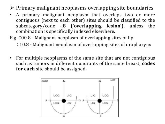
A primary malignant neoplasm that overlaps two or more contiguous (next to each other) sites should be classified to the subcategory/code .8 ('overlapping lesion'), unless the combination is specifically indexed elsewhere.
What chapter is neoplasms classified in?
All neoplasms are classified in this chapter, whether they are functionally active or not. An additional code from Chapter 4 may be used, to identify functional activity associated with any neoplasm. Morphology [Histology] Chapter 2 classifies neoplasms primarily by site (topography), with broad groupings for behavior, malignant, in situ, benign, ...
What is the diagnosis for D04.7?
D04.7 Carcinoma in situ of skin of lower limb, including hip. D04.70 Carcinoma in situ of skin of unspecified lower limb, including hip. D04.71 Carcinoma in situ of skin of right lower limb, including hip. D04.72 Carcinoma in situ of skin of left lower limb, including hip.
What is the table of neoplasms used for?
The Table of Neoplasms should be used to identify the correct topography code. In a few cases, such as for malignant melanoma and certain neuroendocrine tumors, the morphology (histologic type) is included in the category and codes. Primary malignant neoplasms overlapping site boundaries.
What is stage 0 breast cancer?
There are 2 types of stage 0 breast carcinoma in situ: ductal carcinoma in situ (dcis) and lobular carcinoma in situ (lcis). Dcis is a noninvasive condition in which abnormal cells are found in the lining of a breast duct (a tube that carries milk to the nipple).
What is LCis in breast?
Lcis is a condition in which abnormal cells are found in the lobules (small sections of tissue involved with making milk) of the breast. This condition seldom becomes invasive cancer; however, having lcis in one breast increases the risk of developing breast cancer in either breast.
What does "type 1 excludes note" mean?
It means "not coded here". A type 1 excludes note indicates that the code excluded should never be used at the same time as D05. A type 1 excludes note is for used for when two conditions cannot occur together , such as a congenital form versus an acquired form of the same condition. carcinoma in situ of skin of breast (.
What is lobular carcinoma in situ?
Lobular carcinoma in situ (LCIS) is a condition caused by unusual cells in the lobules of the breast. Specialty: Oncology. Diagram showing localized and invasive LCIS. Source: Wikipedia.
What is the ICD code for acute care?
D05.0. Non-Billable means the code is not sufficient justification for admission to an acute care hospital when used a principal diagnosis. Use a child code to capture more detail. ICD Code D05.0 is a non-billable code.
What is the code for a primary malignant neoplasm?
A primary malignant neoplasm that overlaps two or more contiguous (next to each other) sites should be classified to the subcategory/code .8 ('overlapping lesion'), unless the combination is specifically indexed elsewhere.
What is the table of neoplasms used for?
The Table of Neoplasms should be used to identify the correct topography code. In a few cases, such as for malignant melanoma and certain neuroendocrine tumors, the morphology (histologic type) is included in the category and codes. Primary malignant neoplasms overlapping site boundaries.
Can multiple neoplasms be coded?
For multiple neoplasms of the same site that are not contiguous, such as tumors in different quadrants of the same breast, codes for each site should be assigned. Malignant neoplasm of ectopic tissue. Malignant neoplasms of ectopic tissue are to be coded to the site mentioned, e.g., ectopic pancreatic malignant neoplasms are coded to pancreas, ...

Popular Posts:
- 1. icd 10 cm code for first degree av block
- 2. icd 10 code for degenerative joint disease acromioclavicular joint
- 3. icd 10 code for ghtn
- 4. icd 10 code for inflammatory arthropathy
- 5. what is the icd 10 code for mature mediastinal teratoma
- 6. icd 10 code for hearin loss
- 7. icd 10 code for dccd
- 8. icd-10 code for zombie attack
- 9. icd 9 code for sublesional hemorrhage
- 10. icd-10 code for complication of failed skin graft