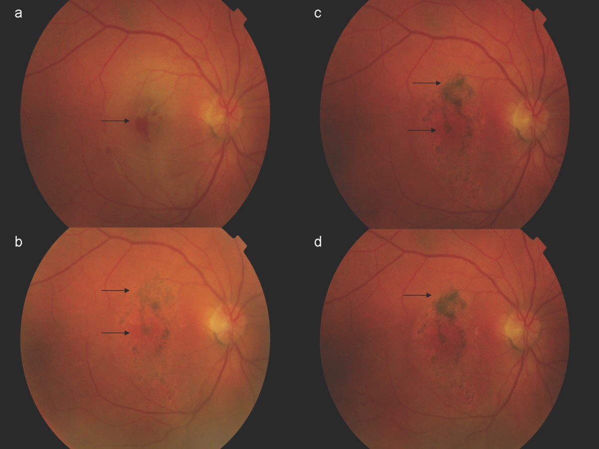
Macular cyst, hole, or pseudohole, right eye
H35. 341 is a billable/specific ICD-10-CM code that can be used to indicate a diagnosis for reimbursement purposes.What is the ICD 10 code for macular cyst with hole?
Oct 01, 2021 · Macular cyst, hole, or pseudohole, right eye. 2016 2017 2018 2019 2020 2021 2022 Billable/Specific Code. H35.341 is a billable/specific ICD-10-CM code that can be used to indicate a diagnosis for reimbursement purposes. The 2022 edition of ICD-10-CM H35.341 became effective on October 1, 2021.
What is the ICD 10 code for pseudohole in left eye?
Oct 01, 2021 · Macular cyst, hole, or pseudohole, right eye Billable Code H35.341 is a valid billable ICD-10 diagnosis code for Macular cyst, hole, or pseudohole, right eye . It is found in the 2022 version of the ICD-10 Clinical Modification (CM) and can be used in all HIPAA-covered transactions from Oct 01, 2021 - Sep 30, 2022 .
What is the ICD 10 code for cysts on the eyelid?
Oct 01, 2021 · Macular cyst, hole, or pseudohole, unspecified eye. H35.349 is a billable/specific ICD-10-CM code that can be used to indicate a diagnosis for reimbursement purposes. The 2022 edition of ICD-10-CM H35.349 became effective on October 1, 2021.
What is the ICD 10 code for pseudohole cyst?
ICD-10 code H35.341 for Macular cyst, hole, or pseudohole, right eye is a medical classification as listed by WHO under the range - Diseases of the eye and adnexa . Subscribe to Codify and get the code details in a flash.
What is macula hole?
A macular hole is a small gap that opens at the centre of the retina, in an area called the macula. The retina is the light-sensitive film at the back of the eye. In the centre is the macula – the part responsible for central and fine-detail vision needed for tasks such as reading.
What is full thickness macular hole?
A Full Thickness Macular Hole (FTMH) is a hole in the retina that occurs in the central part of the macula (known as the fovea).
What is a lamellar hole?
Lamellar macular hole (LMH) is a vitreoretinal disorder characterized by an irregular foveal contour, a break in the inner fovea, dehiscence of the inner foveal retina from the outer retina, and the absence of a full-thickness foveal defect with intact foveal photoreceptors. The pathogenesis is only partially known.Jan 9, 2019
How do you repair a macular hole?
Macular Hole Surgery And Repair A vitrectomy is the most common treatment for macular holes. In this surgery, a retinal specialist removes the vitreous gel to stop it from pulling on the retina. Then the specialist inserts a mixture of air and gas into the space once occupied by the vitreous.
When do you refer a macular hole?
1. Macular hole. When to refer: Once a true retinal break is apparent (stage 2-4), the patient should be referred for surgical treatment. Repair is successful in most patients, and the earlier the treatment, the better the prognosis for the patient.Aug 24, 2015
What is a stage 2 macular hole?
Stages 2–4 include full-thickness macular holes, which are further divided into smaller holes (<400 μm in diameter (stage 2)), holes larger than 400 μm in diameter (stage 3) and with a complete posterior vitreous detachment (stage 4).Oct 23, 2013
What is the difference between macular hole and lamellar hole?
A macular hole is a full thickness defect in the macula whilst a lamellar macular hole is only a partial thicknessdefect in the macula.
What is a macular Pseudohole?
Macular pseudohole: Not a true hole; rather it is a condition in which scar tissue called epiretinal membrane tugs or pulls on the underlying retina, which can look similar to a macular hole during a clinical eye examination.
What is a partial-thickness macular hole?
Partial-thickness (or lamellar) macular hole formation is a lateral or tangential tractional disorder with intact outer retinal layers. Many of these subtle contour changes are asymptomatic but should be observable, and clinicians can initially watch most lamellar holes closely.Oct 15, 2019
Can you go blind from a macular hole?
It's likely you'll have little to no central vision left. If left untreated, these holes can cause serious complications like a detached retina which will also cause problems with your peripheral vision and eventually lead to total blindness.Sep 30, 2019
Do all macular holes require surgery?
Although some macular holes can seal themselves and require no treatment, surgery is necessary in many cases to help improve vision. In this surgical procedure – called a vitrectomy – the vitreous gel is removed to prevent it from pulling on the retina and replaced with a bubble containing a mixture of air and gas.Jul 8, 2019
What is the difference between macular degeneration and a macular hole?
Risk factors for developing age-related macular degeneration include family history, obesity, sleep apnea, smoking, age, and prolonged sun-exposure. A macular hole also involves damage to the macula, however in this case it is caused by age-related changes to the gel-like filling within the eye known as the vitreous.Jan 20, 2016
What does AMD do to the eye?
AMD affects the macula, the part of the eye that allows you to see fine detail. It does not hurt, but it causes cells in the macula to die. There are two types: wet and dry. Wet AMD happens when abnormal blood vessels grow under the macula.
What is AMD in medical terms?
Also called: AMD, Age-related macular degeneration. Macular degeneration, or age-related macular degeneration (AMD), is a leading cause of vision loss in Americans 60 and older. It is a disease that destroys your sharp, central vision.
What is the GEM crosswalk?
The General Equivalency Mapping (GEM) crosswalk indicates an approximate mapping between the ICD-10 code H35.341 its ICD-9 equivalent. The approximate mapping means there is not an exact match between the ICD-10 code and the ICD-9 code and the mapped code is not a precise representation of the original code.
The ICD code H353 is used to code Drusen
Drusen (singular, "druse") are tiny yellow or white accumulations of extracellular material that build up between Bruch's membrane and the retinal pigment epithelium of the eye. The presence of a few small ("hard") drusen is normal with advancing age, and most people over 40 have some hard drusen.
Equivalent ICD-9 Code GENERAL EQUIVALENCE MAPPINGS (GEM)
This is the official approximate match mapping between ICD9 and ICD10, as provided by the General Equivalency mapping crosswalk. This means that while there is no exact mapping between this ICD10 code H35.341 and a single ICD9 code, 362.54 is an approximate match for comparison and conversion purposes.

Popular Posts:
- 1. icd 9 code for leg hematoma
- 2. icd code for basal cell carcinoma
- 3. icd 10 code for chronic aspiration unspecified
- 4. icd 10 code for gouty arthropy
- 5. icd-10 code for superior mediastinal and cervical lymphadenopathy
- 6. icd 10 code for medicine refill
- 7. icd 10 code for anaphylactic shock initial encounter
- 8. icd 10 code for rhinitis medicamentosa
- 9. icd-10 code for cmc fracture dislocation
- 10. icd 10 code for anemia of chronic disease