Other seborrheic keratosis. L82.1 is a billable/specific ICD-10-CM code that can be used to indicate a diagnosis for reimbursement purposes. The 2019 edition of ICD-10-CM L82.1 became effective on October 1, 2018.
L82
What is the ICD 10 code for seborrheic keratosis?
2022 ICD-10-CM Diagnosis Code L82 2022 ICD-10-CM Diagnosis Code L82 Seborrheic keratosis 2016 2017 2018 2019 2020 2021 2022 Non-Billable/Non-Specific Code L82 should not be used for reimbursement purposes as there are multiple codes below it …
What is the ICD 10 code for macular keratitis?
Oct 01, 2021 · L82.1 is a billable/specific ICD-10-CM code that can be used to indicate a diagnosis for reimbursement purposes. The 2022 edition of ICD-10-CM L82.1 became effective on October 1, 2021. This is the American ICD-10-CM version of L82.1 - other international versions of ICD-10 L82.1 may differ. Applicable To Seborrheic keratosis NOS
What is the ICD 10 code for actinic keratosis?
Oct 01, 2021 · 2022 ICD-10-CM Diagnosis Code L85.1 Acquired keratosis [keratoderma] palmaris et plantaris 2016 2017 2018 2019 2020 2021 2022 Billable/Specific Code L85.1 is a billable/specific ICD-10-CM code that can be used to indicate a diagnosis for reimbursement purposes. The 2022 edition of ICD-10-CM L85.1 became effective on October 1, 2021.
What is the ICD 10 code for keratoderma palmaris et plantaris?
Sep 28, 2021 · ICD coding. ICD-10: L82 - seborrheic keratosis Epidemiology. Most common benign skin tumor Age > 50; incidence increases with age M = F ... Macular. Videos. Seborrheic keratosis overview. Seborrheic keratosis and variants. Sample pathology report. …
What is the ICD-10 code for seborrheic keratosis?
L82.0L82. 0 is a billable/specific ICD-10-CM code that can be used to indicate a diagnosis for reimbursement purposes.
What is the meaning of seborrheic keratosis?
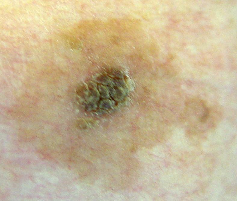
A seborrheic keratosis (seb-o-REE-ik ker-uh-TOE-sis) is a common noncancerous (benign) skin growth. People tend to get more of them as they get older. Seborrheic keratoses are usually brown, black or light tan. The growths (lesions) look waxy or scaly and slightly raised.Jan 18, 2022
What is the CPT code for seborrheic keratosis?
The treatment of common warts, plantar warts, actinic keratosis and seborrheic keratosis by most methods (application of acid, freezing, laser or electrocautery) is covered by “destruction” codes. Use 17000 for destruction of the first lesion. Use add-on code 17003 for each lesion between two and 14.
Is seborrheic keratosis a Macule?
Seborrheic keratosis is caused by the benign proliferation of immature keratinocytes, resulting in well-demarcated, round or oval, flat-shaped macules. They are typically slow-growing, can increase in thickness over time, and they rarely resolve spontaneously.Feb 9, 2022
What is Dermatofibrosis?
Dermatofibromas are small, noncancerous (benign) skin growths that can develop anywhere on the body but most often appear on the lower legs, upper arms or upper back. These nodules are common in adults but are rare in children. They can be pink, gray, red or brown in color and may change color over the years.Jan 9, 2019
What are the different types of keratosis?
More specifically, it can refer to:actinic keratosis (also known as solar keratosis), a premalignant condition.chronic scar keratosis.hydrocarbon keratosis.keratosis pilaris (KP, also known as follicular keratosis)seborrheic keratosis, not premalignant.
What is the ICD 10 code for keratosis?
L85.1Acquired keratosis [keratoderma] palmaris et plantaris L85. 1 is a billable/specific ICD-10-CM code that can be used to indicate a diagnosis for reimbursement purposes.
What does CPT code 17000 mean?
CPT® Code 17000 in section: Destruction (eg, laser surgery, electrosurgery, cryosurgery, chemosurgery, surgical curettement), premalignant lesions (eg, actinic keratoses)
Are seborrheic keratoses premalignant?
Seborrhoeic keratoses are not premalignant tumours. However: Skin cancers are sometimes difficult to tell apart from seborrhoeic keratoses. Skin cancer may by chance arise within or collide with a seborrhoeic keratosis.
Is seborrheic keratosis benign or premalignant?
Seborrheic keratosis is a common benign (noncancerous) skin growth. It tends to appear in middle age and you may get more as you get older. Seborrheic keratoses are not pre-cancerous, but they can resemble other skin growths that are.Aug 27, 2021
Is Verrucous keratosis the same as seborrheic keratosis?
Seborrheic keratoses are common verrucous or stuck-on epidermal papules of various colors (Fig. 448-8). They are commonly seen with advancing age but may arise suddenly (sign of Leser-Trélat) in association with internal malignancy.
How is seborrheic keratosis diagnosed?
Your doctor can usually tell whether you have a seborrheic keratosis by looking at the affected skin. If there is a question about the diagnosis, your doctor might recommend removing the growth so that it can be examined under a microscope.Jan 18, 2022
Popular Posts:
- 1. icd 10 code for obstructive uropathy due to bph
- 2. icd-10 code for rapid ventricular response
- 3. icd 10 code for abnormal vision screening
- 4. icd-10 code for tonsilitis
- 5. icd 9 code for school failure
- 6. icd 10 code for vaginal can
- 7. icd code for preoperative clearance
- 8. icd code for afib with rvr
- 9. icd 10 code for lumbar neurogenic claudication
- 10. icd 10 code for aftercare following ventral hernia repair