Hydatidiform mole, unspecified
O01. 9 is a billable/specific ICD-10-CM code that can be used to indicate a diagnosis for reimbursement purposes. The 2022 edition of ICD-10-CM O01. 9 became effective on October 1, 2021.Is a hydatidiform mole cancerous?
A hydatidiform mole contains many cysts (sacs of fluid). It is usually benign (not cancer) but it may spread to nearby tissues (invasive mole). It may also become a malignant tumor called choriocarcinoma. Hydatidiform mole is the most common type of gestational trophoblastic tumor.
What is hydatidiform mole O01?
Hydatidiform mole O01- >. A slow-growing tumor that develops from trophoblastic cells (cells that help an embryo attach to the uterus and help form the placenta) after fertilization of an egg by a sperm. A hydatidiform mole contains many cysts (sacs of fluid). It is usually benign (not cancer) but it may spread to nearby tissues (invasive mole).
What is the incidence of choriocarcinoma with hydatidiform mole?
The incidence of choriocarcinoma is higher in patients with complete hydatidiform mole. When a hydatidiform mole invades the myometrium and broad ligament, or it is found in distant sites as vagina, vulva, and lung, it is referred as invasive mole.
What is a hydatidiform tumor?
A slow-growing tumor that develops from trophoblastic cells (cells that help an embryo attach to the uterus and help form the placenta) after fertilization of an egg by a sperm. A hydatidiform mole contains many cysts (sacs of fluid). It is usually benign (not cancer) but it may spread to nearby tissues (invasive mole).
What is CPT code for hydatidiform mole?
ICD-10-CM Code for Hydatidiform mole, unspecified O01. 9.
What hydatidiform mole means?
Listen to pronunciation. (HY-duh-TIH-dih-form...) A slow-growing tumor that develops from trophoblastic cells (cells that help an embryo attach to the uterus and help form the placenta) after fertilization of an egg by a sperm. A hydatidiform mole contains many cysts (sacs of fluid).
Is hydatidiform mole malignant?
A hydatidiform mole is considered malignant if metastases or destructive invasion of the myometrium (ie, invasive mole) occurs, or when the serum hCG levels plateau or rise during the period of follow-up and an intervening pregnancy is excluded.
Where is a hydatidiform mole?
Hydatidiform mole (HM) is a rare mass or growth that forms inside the womb (uterus) at the beginning of a pregnancy. It is a type of gestational trophoblastic disease (GTD).
Is molar pregnancy and hydatidiform mole same?
A molar pregnancy — also known as hydatidiform mole — is a rare complication of pregnancy characterized by the abnormal growth of trophoblasts, the cells that normally develop into the placenta. There are two types of molar pregnancy, complete molar pregnancy and partial molar pregnancy.
How is hydatidiform mole diagnosis?
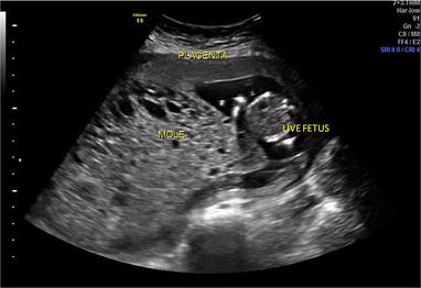
Ultrasonography is done to be sure that the growth is a hydatidiform mole and not a fetus or amniotic sac (which contains the fetus and fluid around it). (D and C) or obtained when tissue is passed and is then examined under a microscope (biopsy) to confirm the diagnosis.
What causes hydatidiform moles?
When a normal sperm cell fertilizes one of these oocytes, the resulting embryo has only one set of chromosomes. Because the embryo has no genes from the mother, the pregnancy cannot develop normally, resulting in a hydatidiform mole.
Is vesicular mole and hydatidiform mole same?
Based on morphology, hydatidiform moles can be divided into two types: in complete moles, all the chorionic villi are vesicular, and no sign of embryonic or fetal development is present.
How do you say Hydatidiform?
Phonetic spelling of hydatidiform molehy-datidi-form mole. Abner Lynch.hydatidiform mole. Dashawn Olson.hy-da-tid-i-form mole.
What is the difference between a complete and incomplete hydatidiform mole?
In complete hydatidiform mole, there is no fetal tissue present; in partial hydatiform moles, there is some residual fetal tissue. Both are due to the over-proliferation of chorionic villi.
What is pathognomonic molar pregnancy?
The most common presenting symptom of molar pregnancy is vaginal bleeding, reported in up to 97% of patients. Intrauterine clots may undergo oxidation and liquefaction, producing pathognomonic prune juice-like fluid. Rarely, spontaneous expulsion of grape-like villi will provide the diagnosis of hydatidiform mole.
When are moles usually present?
Complete moles are usually diploid and typically present between the eleventh and twenty-fifth week of pregnancy, whereas partial moles are usually triploid and usually present around the nineteenth week of pregnancy. The incidence of choriocarcinoma is higher in patients with complete hydatidiform mole.
What is the most common type of gestational trophoblastic tumor?
It may also become a malignant tumor called choriocarcinoma. Hydatidiform mole is the most common type of gestational trophoblastic tumor. Trophoblastic hyperplasia associated with normal gestation, or molar pregnancy. It is characterized by the swelling of the chorionic villi and elevated human chorionic gonadotropin.
What does the title of a manifestation code mean?
In most cases the manifestation codes will have in the code title, "in diseases classified elsewhere.". Codes with this title are a component of the etiology/manifestation convention. The code title indicates that it is a manifestation code.
What does "type 1 excludes note" mean?
A type 1 excludes note is for used for when two conditions cannot occur together, such as a congenital form versus an acquired form of the same condition. chorioadenoma (destruens) (.
What is a partial mole?
Partial moles are characterized by a mixture of large hydropic villi and normal placenta tissue. Complete moles are usually diploid and typically present between the eleventh and twenty-fifth week of pregnancy, whereas partial moles are usually triploid and usually present around the nineteenth week of pregnancy.
What is the most common type of gestational trophoblastic tumor?
It may also become a malignant tumor called choriocarcinoma. Hydatidiform mole is the most common type of gestational trophoblastic tumor. Trophoblastic hyperplasia associated with normal gestation, or molar pregnancy. It is characterized by the swelling of the chorionic villi and elevated human chorionic gonadotropin.
What does the title of a manifestation code mean?
In most cases the manifestation codes will have in the code title, "in diseases classified elsewhere.". Codes with this title are a component of the etiology/manifestation convention. The code title indicates that it is a manifestation code.
What does "type 1 excludes note" mean?
A type 1 excludes note is for used for when two conditions cannot occur together, such as a congenital form versus an acquired form of the same condition. chorioadenoma (destruens) (.
What is a hydatidiform mole?
According to the amount of villous involvement, a hydatidiform mole is defined as complete or partial.
What is the karyotype of a mole?
Most complete moles (>90%) have a 46,xx karyotype and the rest 46,xy karyotype. Trophoblastic hyperplasia associated with normal gestation, or molar pregnancy. It is characterized by the swelling of the chorionic villi and elevated human chorionic gonadotropin.
What is the rare cancer in women of childbearing age?
A rare cancer in women of childbearing age in which cancer cells grow in the tissues that are formed in the uterus after conception. Also called gestational trophoblastic disease, gestational trophoblastic neoplasia, gestational trophoblastic tumor, or choriocarcinoma.
When are moles usually present?
Complete moles are usually diploid and typically present between the eleventh and twenty-fifth week of pregnancy, whereas partial moles are usually triploid and usually present around the nineteenth week of pregnancy. The incidence of choriocarcinoma is higher in patients with complete hydatidiform mole.
Is a hydatidiform mole a tumor?
It may also become a malignant tumor called choriocarcinoma. Hydatidiform mole is the most common type of gestational trophoblastic tumor.

Popular Posts:
- 1. what's the icd 10 code for . systemic lupus erythematosus with lung involvement
- 2. icd-10-cm code for abscess right finger
- 3. icd 10 code for closed fracture of multiple ribs right side
- 4. icd 10 cm code for acute and chronic sinusitis
- 5. icd-10-cm code for tension headache
- 6. what is the correct icd 10 code for d64
- 7. icd 10 code for scc knee
- 8. icd 10 cm code for lump in chest
- 9. icd 10 code for axillary abscess
- 10. icd 10 code for bulging fontanelle