Melanocytic nevi of unspecified part of face
D22. 30 is a billable/specific ICD-10-CM code that can be used to indicate a diagnosis for reimbursement purposes. The 2022 edition of ICD-10-CM D22. 30 became effective on October 1, 2021.What is the ICD 10 code for nevus?
I78.1 is a valid billable ICD-10 diagnosis code for Nevus, non-neoplastic . It is found in the 2022 version of the ICD-10 Clinical Modification (CM) and can be used in all HIPAA-covered transactions from Oct 01, 2021 - Sep 30, 2022 .
What is the ICD 10 code for melanocytic nevi?
Melanocytic nevi of other parts of face. D22.39 is a billable/specific ICD-10-CM code that can be used to indicate a diagnosis for reimbursement purposes. The 2020 edition of ICD-10-CM D22.39 became effective on October 1, 2019.
What is the ICD 10 code for uveitis?
Q82.5 is a billable/specific ICD-10-CM code that can be used to indicate a diagnosis for reimbursement purposes. The 2018/2019 edition of ICD-10-CM Q82.5 became effective on October 1, 2018. This is the American ICD-10-CM version of Q82.5 - other international versions of ICD-10 Q82.5 may differ.
What is the ICD 10 code for Neurologic diagnosis?
D22.30 is a billable/specific ICD-10-CM code that can be used to indicate a diagnosis for reimbursement purposes. The 2018/2019 edition of ICD-10-CM D22.30 became effective on October 1, 2018. This is the American ICD-10-CM version of D22.30 - other international versions of ICD-10 D22.30 may differ.
What is the code for a primary malignant neoplasm?
What chapter is neoplasms classified in?
About this website
What is the ICD-10 code for nevus?
I78.11.
What is melanocytic nevi of unspecified part of face?
Melanocytic nevi are benign neoplasms or hamartomas composed of melanocytes, the pigment-producing cells that constitutively colonize the epidermis.
What is an intradermal nevus?
Intradermal melanocytic nevi are common, benign, pigmented skin tumors formed by proliferation of dermal melanocytes. A number of notable, uncommon changes may be observed in intradermal melanocytic nevi. In particular, their association with lymphatic invasion is an extremely rare phenomenon.
What is code D22 39?
ICD-10 Code for Melanocytic nevi of other parts of face- D22. 39- Codify by AAPC.
Is a melanocytic nevus a mole?
Melanocytic naevi are pigmented moles. The word 'melanocytic' means that they are made up of the cells (melanocytes) which produce the dark pigment (melanin) that gives the skin its colour. Melanocytes clustered together form naevi.
Is a nevus a mole?
Most people continue to develop new moles until about age 40. In older people, common moles tend to fade away. Another name for a mole is a nevus. The plural is nevi.
What is an atypical nevus?
Atypical nevi, also known as dysplastic nevi, are benign acquired melanocytic neoplasms. Atypical nevi share some of the clinical features of melanoma, such as asymmetry, irregular borders, multiple colors, and diameter >5 mm (picture 1A). They occur sporadically or in a familial setting.
What is a complex nevus?
A compound naevus is a normal and harmless group of melanocytes (pigment cells) from different skin layers, and may be irregular in shape, colour and elevation.
What are 4 types of moles?
There are 4 common types of moles: congenital moles, dysplastic nevi, acquired nevi, and spitz nevi. Below are the differences between each.
Is a compound nevus benign or malignant?
benignCompound Nevi Typically they are light tan to dark brown, dome shaped papules that are 1-10 mm in diameter. Compound Nevi are benign proliferations of melanocytes at the epidermal-dermal junction.
What is the ICD-10 code for skin lesion?
ICD-10-CM Code for Disorder of the skin and subcutaneous tissue, unspecified L98. 9.
What causes intradermal nevus?
sun damage, especially for those with fairer skin. immunosuppressive treatments, such as those used in cancer, which can cause more moles to develop. genetic factors, such as your parents having a lot of moles, which makes it more likely that you will have them as too.
Does melanocytic mean melanoma?
Melanocytes: These are the cells that can become melanoma. They normally make a brown pigment called melanin, which gives the skin its tan or brown color.
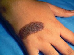
What does a melanocytic nevi look like?
They typically appear as small brown, tan, or pink spots. You can be born with moles or develop them later. Moles that you're born with are known as congenital moles. However, most moles develop during childhood and adolescence.
Should melanocytic nevus be removed?
to the editor: In the article on newborn skin, the authors recommend removal of large and giant congenital melanocytic nevi as the current management strategy. In fact, complete nevus removal is impossible for many large nevi and virtually all giant nevi.
What causes melanocytic nevus?
Congenital melanocytic nevi are caused by a change in color (pigment) cells of the skin. The moles happen by chance. CMN is not passed down from the parents. There is no way to prevent your child from being born with moles.
What is the code for a primary malignant neoplasm?
A primary malignant neoplasm that overlaps two or more contiguous (next to each other) sites should be classified to the subcategory/code .8 ('overlapping lesion'), unless the combination is specifically indexed elsewhere.
What chapter is neoplasms classified in?
All neoplasms are classified in this chapter, whether they are functionally active or not. An additional code from Chapter 4 may be used, to identify functional activity associated with any neoplasm. Morphology [Histology] Chapter 2 classifies neoplasms primarily by site (topography), with broad groupings for behavior, malignant, in situ, benign, ...
What is the plural of "nevus"?
The plural of nevus is nevi (nee-vye). A benign (not cancer) growth on the skin that is formed by a cluster of melanocytes (cells that make a substance called melanin, which gives color to skin and eyes). A mole is usually dark and may be raised from the skin.
What is the code for a primary malignant neoplasm?
A primary malignant neoplasm that overlaps two or more contiguous (next to each other) sites should be classified to the subcategory/code .8 ('overlapping lesion'), unless the combination is specifically indexed elsewhere.
What is a benign growth on the skin?
A benign growth on the skin (usually tan, brown, or flesh-colored) that contain s a cluster of melanocytes and surrounding supportive tissue. A neoplasm composed of melanocytes that usually appears as a dark spot on the skin. A nevus characterised by the presence of excessive pigment. A nevus containing melanin.
What is the code for a primary malignant neoplasm?
A primary malignant neoplasm that overlaps two or more contiguous (next to each other) sites should be classified to the subcategory/code .8 ('overlapping lesion'), unless the combination is specifically indexed elsewhere.
What chapter is neoplasms classified in?
All neoplasms are classified in this chapter, whether they are functionally active or not. An additional code from Chapter 4 may be used, to identify functional activity associated with any neoplasm. Morphology [Histology] Chapter 2 classifies neoplasms primarily by site (topography), with broad groupings for behavior, malignant, in situ, benign, ...
What is the ICd 10 code for Nevus?
I78.1 is a valid billable ICD-10 diagnosis code for Nevus, non-neoplastic . It is found in the 2021 version of the ICD-10 Clinical Modification (CM) and can be used in all HIPAA-covered transactions from Oct 01, 2020 - Sep 30, 2021 .
What does NEC not elsewhere mean?
NEC Not elsewhere classifiable#N#This abbreviation in the Tabular List represents “other specified”. When a specific code is not available for a condition, the Tabular List includes an NEC entry under a code to identify the code as the “other specified” code.
Do you include decimal points in ICD-10?
DO NOT include the decimal point when electronically filing claims as it may be rejected. Some clearinghouses may remove it for you but to avoid having a rejected claim due to an invalid ICD-10 code, do not include the decimal point when submitting claims electronically. See also: Angioma see also Hemangioma, by site.
What is the code for a primary malignant neoplasm?
A primary malignant neoplasm that overlaps two or more contiguous (next to each other) sites should be classified to the subcategory/code .8 ('overlapping lesion'), unless the combination is specifically indexed elsewhere.
What chapter is neoplasms classified in?
All neoplasms are classified in this chapter, whether they are functionally active or not. An additional code from Chapter 4 may be used, to identify functional activity associated with any neoplasm. Morphology [Histology] Chapter 2 classifies neoplasms primarily by site (topography), with broad groupings for behavior, malignant, in situ, benign, ...
Can multiple neoplasms be coded?
For multiple neoplasms of the same site that are not contiguous, such as tumors in different quadrants of the same breast, codes for each site should be assigned. Malignant neoplasm of ectopic tissue. Malignant neoplasms of ectopic tissue are to be coded to the site mentioned, e.g., ectopic pancreatic malignant neoplasms are coded to pancreas, ...
What is the code for a primary malignant neoplasm?
A primary malignant neoplasm that overlaps two or more contiguous (next to each other) sites should be classified to the subcategory/code .8 ('overlapping lesion'), unless the combination is specifically indexed elsewhere.
What chapter is neoplasms classified in?
All neoplasms are classified in this chapter, whether they are functionally active or not. An additional code from Chapter 4 may be used, to identify functional activity associated with any neoplasm. Morphology [Histology] Chapter 2 classifies neoplasms primarily by site (topography), with broad groupings for behavior, malignant, in situ, benign, ...
Popular Posts:
- 1. icd 10 code for blindness due to hypertension
- 2. icd-9 code for external cause place of occurence home
- 3. icd 9 code for aortic aneurysm
- 4. icd 9 code for pulmonary infiltrates
- 5. icd 10 code for failed swallow study
- 6. icd 10 code for shift work
- 7. icd 9 code for epigastric abdominal pain
- 8. icd 10 code for odontogenic infection
- 9. icd 10 code for rll cellulitis
- 10. icd-10-pcs 2017 code for right ovarian cystectomy