133.
What is the ICD 10 code for blindness to the left eye?
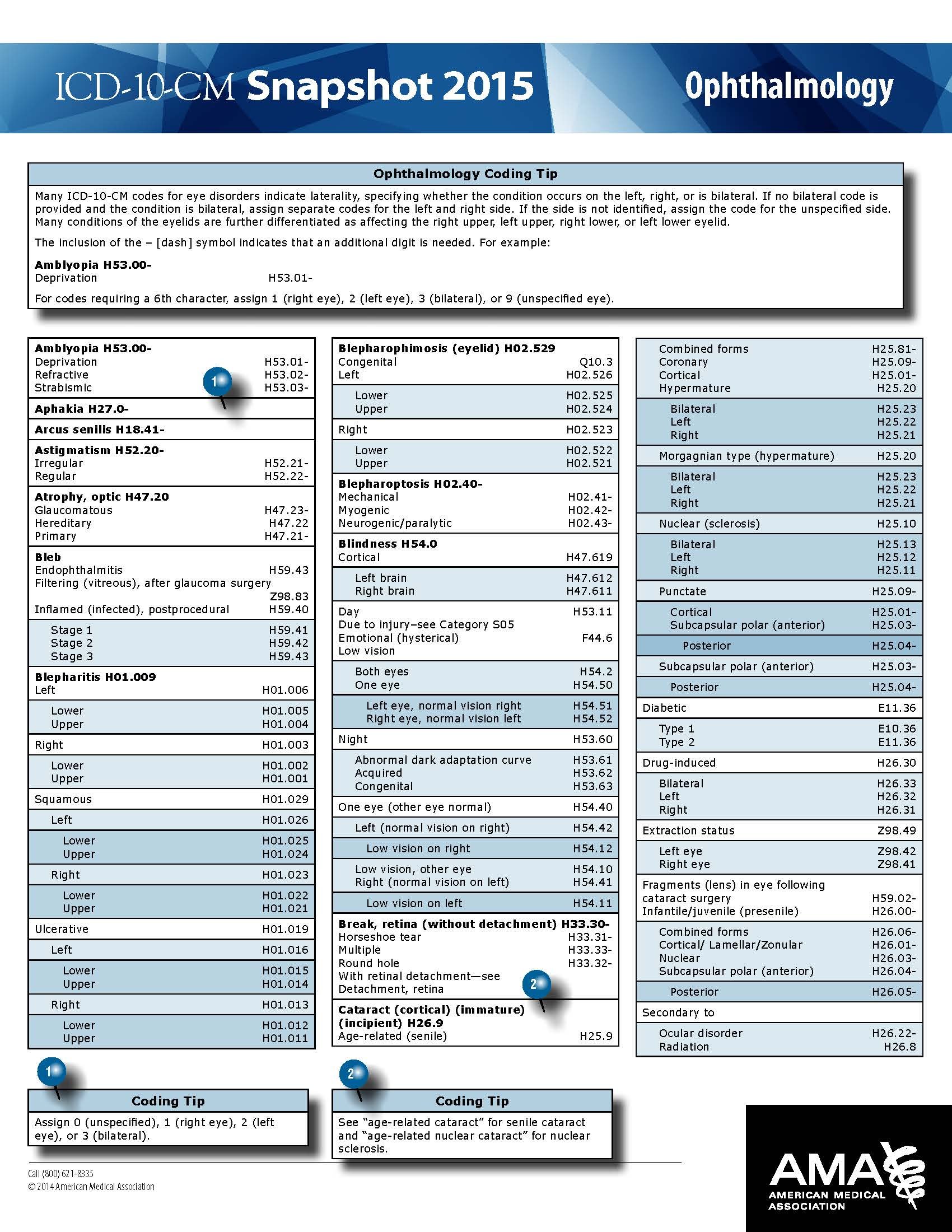
H54.42 ICD-10 code on the other hand will be used to represent blindness to the left eye accompanied with normal vision to the right eye. H54.50 will be used to represent low vision on one eye accompanied with unspecified eye.
What is the ICD 10 code for pterygium of left eye?
2018/2019 ICD-10-CM Diagnosis Code H11.002. Unspecified pterygium of left eye. H11.002 is a billable/specific ICD-10-CM code that can be used to indicate a diagnosis for reimbursement purposes.
What is the ICD 10 code for trauma to the eye?
H21.232 is a billable/specific ICD-10-CM code that can be used to indicate a diagnosis for reimbursement purposes. The 2022 edition of ICD-10-CM H21.232 became effective on October 1, 2021. This is the American ICD-10-CM version of H21.232 - other international versions of ICD-10 H21.232 may differ. injury (trauma) of eye and orbit ( S05.-)
What is the ICD 10 code for cataract surgery?
ICD-10 code Z98.42 for Cataract extraction status, left eye is a medical classification as listed by WHO under the range - Factors influencing health status and contact with health services . Subscribe to Codify and get the code details in a flash.
What is the diagnosis code for cataract left eye?
ICD-10 Code for Combined forms of age-related cataract, left eye- H25. 812- Codify by AAPC.
What is the ICD-10 diagnosis code for posterior vitreous detachment left eye?
ICD-10 code H43. 812 for Vitreous degeneration, left eye is a medical classification as listed by WHO under the range - Diseases of the eye and adnexa .
What is the ICD-10 code for small pupil left eye?
Pupillary abnormality, unspecified eye H21. 569 is a billable/specific ICD-10-CM code that can be used to indicate a diagnosis for reimbursement purposes. The 2022 edition of ICD-10-CM H21. 569 became effective on October 1, 2021.
Is pigmentary glaucoma open-angle?
Pigmentary glaucoma is a type of secondary open-angle glaucoma characterized by heavy homogenous pigmentation of the trabecular meshwork, iris transillumination defects, and pigment along the corneal endothelium (Krukenberg spindle).
What is posterior vitreous detachment?
Posterior vitreous detachment (PVD) occurs when the gel that fills the eyeball separates from the retina. It's a natural, normal part of aging. PVD can cause floaters or flashes in your sight, which usually become less noticeable over time. The condition isn't painful, and it doesn't cause vision loss on its own.
How is posterior vitreous detachment diagnosis?
Diagnostic testing Posterior vitreous detachment is usually diagnosed with a dilated eye examination. However, if the vitreous gel is very clear, it may be hard to see the PVD without additional testing, such as optical coherence tomography (OCT) or ocular ultrasound (see Figure 2).
What is the medical term for unequal pupils?
Uneven pupil size, or anisocoria, may be a normal variation in a person's eyes or may indicate an underlying problem.
What does it mean when your pupil isn't centered?
People with Axenfeld-Rieger syndrome often have a pupil that is off-center (corectopia) or extra holes in the iris that can look like multiple pupils (polycoria). This condition can also cause abnormalities of the cornea, which is the clear front covering of the eye.
What does unequal pupils mean?
Unequal pupil sizes of more than 1 mm that develop later in life and do not return to equal size may be a sign of an eye, brain, blood vessel, or nerve disease.
What is open-angle glaucoma?
Open-angle glaucoma is the most common form of the disease. The drainage angle formed by the cornea and iris remains open, but the trabecular meshwork is partially blocked. This causes pressure in the eye to gradually increase. This pressure damages the optic nerve.
What are the two types of glaucoma?
Although there are many types of glaucoma, ophthalmologists typically group them into two main categories: open-angle glaucoma and angle-closure glaucoma. Forms of glaucoma in both categories are characterized by damage to the optic nerve which can eventually lead to blindness.
How is primary open angle glaucoma diagnosed?
Diagnosis is by ophthalmoscopy, gonioscopy, visual field examination, and measurement of central corneal thickness and IOP. Treatment includes topical drugs (eg, prostaglandin analogs, beta-blockers) and often requires laser or incisional surgery to increase aqueous drainage.
Convert 08Q1XZZ to ICD-9-PCS
The following crosswalk between ICD-10-PCS to ICD-9-PCS is based based on the General Equivalence Mappings (GEMS) information:
What is ICD-10-PCS?
The ICD-10 Procedure Coding System (ICD-10-PCS) is a catalog of procedural codes used by medical professionals for hospital inpatient healthcare settings. The Centers for Medicare and Medicaid Services (CMS) maintain the catalog in the U.S. releasing yearly updates.
ICD 10 Eye Glaucoma Code (H40.9)
Glaucoma is an eye condition that results in the optic nerve of the eye worsening with time. This condition is normally associated with increase in the buildup of pressure in the eye. H40.9 is the ICD code that has been designated for this eye condition.
ICD 10 Cataract Unspecified Code (H25.9)
Cataracts is a common eye condition that is known to be a major cause of blindness in many people. Cataract is the clouding of the lens inside the eye which normally results in reduced vision. H25.9 is an ICD 10 code that specifies unspecified age related cataract.
Blindness and Low Vision ICD 10 Codes
Low Vision is a term used to refer to a significant reduction of visual function that cannot be fully corrected by ordinary glasses, contact lenses or any sort of medical treatment. Level of vision codes will also be predominant in ICD10; the only change is that ICD-10 will feature 17 codes in this case as compared to 16 in ICD-9.
Popular Posts:
- 1. icd 10 code for contusion scalp
- 2. icd 9 code for family history of factor v leiden
- 3. icd code for sprained ankle
- 4. icd 10 code for akl 2 dehydration
- 5. what is the icd-10 code for 410
- 6. icd 10 code for epididymo orchitis
- 7. icd 10 code for basal cell carcinoma, right ear?
- 8. icd 10 code for ventricular premature contructure
- 9. icd 10 code for bite by human
- 10. icd 10 code for diasobic chf