Abnormal electrocardiogram [ECG] [EKG]
R94. 31 is a billable/specific ICD-10-CM code that can be used to indicate a diagnosis for reimbursement purposes. The 2022 edition of ICD-10-CM R94. 31 became effective on October 1, 2021.What is poor R wave progression on ECG?
Oct 01, 2021 · R94.31 is a billable/specific ICD-10-CM code that can be used to indicate a diagnosis for reimbursement purposes. The 2022 edition of ICD-10-CM R94.31 became effective on October 1, 2021. This is the American ICD-10-CM version of R94.31 - other international versions of ICD-10 R94.31 may differ. Type 1 Excludes long QT syndrome ( I45.81)
What is the ICD 10 code for abnormal ECG?
Oct 01, 2021 · 2022 ICD-10-CM Diagnosis Code R46.4 2022 ICD-10-CM Diagnosis Code R46.4 Slowness and poor responsiveness 2016 2017 2018 2019 2020 2021 2022 Billable/Specific Code R46.4 is a billable/specific ICD-10-CM code that can be used to indicate a diagnosis for reimbursement purposes. The 2022 edition of ICD-10-CM R46.4 became effective on October …
What is poor R-wave progression (PRWP)?
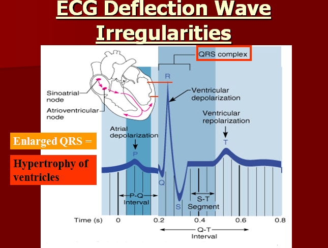
Abstract. Poor R-wave progression is a common ECG finding that is often inconclusively interpreted as suggestive, but not diagnostic, of anterior myocardial infarction (AMI). Recent studies have shown that poor R-wave progression has the following four distinct major causes: AMI, left ventricular hypertrophy, right ventricular hypertrophy, and a variant of normal with …
What does PRWP stand for on ECG?
R94.31 is a billable diagnosis code used to specify a medical diagnosis of abnormal electrocardiogram [ecg] [ekg]. The code R94.31 is valid during the fiscal year 2022 from October 01, 2021 through September 30, 2022 for the submission of HIPAA-covered transactions. The ICD-10-CM code R94.31 might also be used to specify conditions or terms like ambulatory ecg …

What does R94 31 mean?
Abnormal electrocardiogramICD-10 code R94. 31 for Abnormal electrocardiogram [ECG] [EKG] is a medical classification as listed by WHO under the range - Symptoms, signs and abnormal clinical and laboratory findings, not elsewhere classified .
What is the ICD-10 code for borderline ECG?
R94.31R94. 31 - Abnormal electrocardiogram [ECG] [EKG]. ICD-10-CM.
What diagnosis code covers EKG?
Electrocardiogram (ECG or EKG) – CPT 93000, 93005, 93010 – ICD 10 CODE R94.
What is the ICD-10 code for prolonged QTC?
81.
What is borderline ECG?
Dr. Bipin Ninan Abraham. General Physician 7 yrs exp Bangalore. I can't give you a specific diagnosis without seeing the ECG, but it usually means that the findings are within normal limits but closer to being abnormal.Sep 26, 2017
What is the ICD-10 code for abnormal EKG?
R94.31Abnormal electrocardiogram [ECG] [EKG] R94. 31 is a billable/specific ICD-10-CM code that can be used to indicate a diagnosis for reimbursement purposes.
Is ECG and EKG the same?
An electrocardiogram records the electrical signals in the heart. It's a common and painless test used to quickly detect heart problems and monitor the heart's health. An electrocardiogram — also called ECG or EKG — is often done in a health care provider's office, a clinic or a hospital room.Mar 19, 2022
What is the CPT code for ECG?
Rhythm ECGs are used to evaluate signs and symptoms that may reflect a cardiac rhythm disorder. A rhythm ECG interpretation and report only (93042) is included in a 12-lead ECG interpretation and report (93000 or 93010). A rhythm ECG tracing (93040 or 93041) is included in a 12-lead ECG tracing (93000 or 93005).
What is the difference between 93005 and 93010?
93005 is the tracing only without interpretation and report and 93010 is the interpretation and report only. We would expect providers to bill global if both the test and interpretation was performed by the same physician.
What is the ICD 10 code for weakness?
R53.1R53. 1 is a billable/specific ICD-10-CM code that can be used to indicate a diagnosis for reimbursement purposes.
What is the ICD 10 code for elevated troponin?
R74.8Elevated Troponin should be coded to R74. 8 Abnormal levels of other serum enzymes. [Effective 11 Jul 2012, ICD-10-AM/ACHI/ACS 7th Ed.]
What is the ICD 10 code for thrombocytosis?
D75.832022 ICD-10-CM Diagnosis Code D75. 83: Thrombocytosis.Oct 1, 2021
What is the code for EKG?
R94.31 is a billable diagnosis code used to specify a medical diagnosis of abnormal electrocardiogram [ecg] [ekg]. The code R94.31 is valid during the fiscal year 2021 from October 01, 2020 through September 30, 2021 for the submission of HIPAA-covered transactions.
What is an EKG?
Electrocardiogram (EKG), (ECG) An electrocardiogram, also called an ECG or EKG, is a painless test that detects and records your heart's electrical activity. It shows how fast your heart is beating and whether its rhythm is steady or irregular. An EKG may be part of a routine exam to screen for heart disease.
What is the purpose of an echocardiogram?
Echocardiography, or echo, is a painless test that uses sound waves to create moving pictures of your heart. The pictures show the size and shape of your heart. They also show how well your heart's chambers and valves are working. Doctors use an echo to diagnose many different heart problems, and to check how severe they are.
What is a type 1 exclude note?
Type 1 Excludes. A type 1 excludes note is a pure excludes note. It means "NOT CODED HERE!". An Excludes1 note indicates that the code excluded should never be used at the same time as the code above the Excludes1 note.
What is a cardiac MRI?
Cardiac MRI (magnetic resonance imaging) is a painless imaging test that uses radio waves, magnets, and a computer to create detailed pictures of your heart. It can help your doctor figure out whether you have heart disease, and if so, how severe it is. A cardiac MRI can also help your doctor decide the best way to treat heart problems such as
How to identify poor R wave progression?
Poor R wave progression can be identified by the timing of the R wave peak. The following ECG tracings show the slowed progression, along with other signs associated with the issue being diagnosed.
When do delta waves occur?
Delta waves occur when the ventricle is activated too early prior to the AV node being activated. Figure 5: Reversed Limb Leads. In some cases, a poor r wave progression can occur from the limb leads being reversed.
What is QRS transition zone?
QRS transition zone is related to the electrical axis of the heart in the horizontal plane and is easily determined from the precordial leads of a standard 12-lead ECG. However, whether delayed QRS transition, or clockwise rotation of the heart, carries prognostic implications and predicts sudden cardiac death (SCD) is unclear.
What are the characteristics of people with early transition occurring before V3?
People with early transition occurring before V 3 were slightly younger, were less likely to smoke, had a slightly lower BMI and blood pressure, and were less likely to have cardiovascular disease or be taking medication than the rest of the subjects ( Table 1 ).
Is delayed QRS a risk marker for SCD?
Delayed QRS transition in the precordial leads of an ECG seems to be a novel ECG risk marker for SCD. In particular, markedly delayed transition was associated with significantly increased risk of SCD, independent of confounding factors.
Popular Posts:
- 1. icd 10 code for urinary accidents
- 2. icd 10 code for stage 2 pressure would to right ear
- 3. icd 10 code for meconium in amniotic fluid
- 4. icd-9 code for titer screening
- 5. what is the correct icd 10 code for diverticulitis with perforation
- 6. icd 9 code for memory deficit
- 7. icd 10 code for cellulitis of lower leg
- 8. icd code for post cabg x 3
- 9. icd 9 code for coagulation defect
- 10. 2016 icd 10 code for tear l5