91 for Basal cell carcinoma of skin, unspecified is a medical classification as listed by WHO under the range - Malignant neoplasms .
Full
AnswerWhat is ICD 10 code covers A1C?
- Hemoglobin A1c (HbA1c) Test for Diabetes
- What is Hemoglobin A1c (HbA1c) Test for Diabetes?
- A Novel Intervention Including Individualized Nutritional Recommendations Reduces Hemoglobin A1c Level, Medication Use, and Weight in Type 2 Diabetes
How to treat your scars after basal cell carcinoma?
- Avoid the sun whenever possible. When you’re outdoors during the day, seek shade and try to avoid being outdoors when the sun is strongest, between 10:00 a.m. ...
- Protect your skin by wearing sunscreen every single day. ...
- Wear clothing that protects your skin from the sun. ...
What are the odds of a basal cell carcinoma metastasizing?
From the Journal of the American Academy of Dermatology (August 2008): Basal cell carcinoma (BCC), the most common human malignancy, metastasizes in 0.0028% to 0.5% of cases, usually to the lymph nodes, lungs, bones, and skin. After metastatic spread of BCC, survival averages 1 to 2 years.
What is differential diagnosis of basal cell carcinoma?
What is the dermoscopic differential diagnosis of basal cell carcinoma? Differential diagnoses for basal cell carcinoma are: Sebaceous hyperplasia; Melanoma; Pilomatricoma; Trichoepithelioma. Dermoscopic differential diagnosis of BCC
What is the ICD-10 code C44 311?
Basal cell carcinoma of skin of noseICD-10 code C44. 311 for Basal cell carcinoma of skin of nose is a medical classification as listed by WHO under the range - Malignant neoplasms .
What is the ICD-10 code C44 319?
Basal cell carcinoma of skin of other partsICD-10 code C44. 319 for Basal cell carcinoma of skin of other parts of face is a medical classification as listed by WHO under the range - Malignant neoplasms .
What is a basal cell carcinoma?
Basal cell carcinoma is a type of skin cancer that most often develops on areas of skin exposed to the sun, such as the face. On brown and Black skin, basal cell carcinoma often looks like a bump that's brown or glossy black and has a rolled border. Basal cell carcinoma is a type of skin cancer.
What are the two types of basal cell carcinomas?
There are four main clinical variants of basal cell carcinoma. These are nodular, superficial spreading, sclerosing and pigmented basal cell carcinomas.
What is the CPT code for excision of basal cell carcinoma?
Article - Billing and Coding: Excision of Malignant Skin Lesions (A57660)
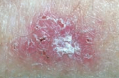
What does basal cell carcinoma look like?
What does BCC look like? BCCs can look like open sores, red patches, pink growths, shiny bumps, scars or growths with slightly elevated, rolled edges and/or a central indentation. At times, BCCs may ooze, crust, itch or bleed. The lesions commonly arise in sun-exposed areas of the body.
Is basal cell carcinoma malignant or benign?
Basal cell carcinoma (BCC) is most often a benign form of skin cancer caused by exposure to ultraviolet (UV) light. However, it's the most frequently occurring form of all skin cancers, with more than 3 million people developing BCC in the U.S. every year.
What is the difference between a basal cell carcinoma and a squamous cell carcinoma?
Basal cell carcinoma accounts for more than 90 percent of all skin cancers in the United States and is the most common of all cancers. Typically, it is a slow-growing cancer that seldom spreads to other parts of the body. Squamous cell carcinoma also rarely spreads, but does so more often than basal cell carcinoma.
How serious is a basal cell carcinoma?
Basal cell cancer is the most common form of skin cancer, accounting for nearly 80% of all skin cancers. Basal cell cancers arise from abnormal basal cells in the skin. It is rarely fatal, but it can be locally aggressive.
What's worse basal cell or squamous?
Though not as common as basal cell (about one million new cases a year), squamous cell is more serious because it is likely to spread (metastasize).
Which is worse basal cell carcinoma or melanoma?
Melanoma is a serious form of skin cancer that begins in cells known as melanocytes. While it is less common than basal cell carcinoma (BCC) and squamous cell carcinoma (SCC), this skin cancer is more dangerous because of its ability to spread to other organs more rapidly if it is not treated at an early stage.
How long does it take for basal cell carcinoma to metastasize?
In one review of 170 cases, the median interval between onset of BCC and metastasis was 9 years, with a range of less than 1 year to 45 years. Despite the long period from onset to metastasis, the tumor behaves aggressively once metastasis occurs.
What is the survival rate for basal cell carcinoma?
The prognosis for patients with BCC is excellent, with a 100% survival rate for cases that have not spread to other sites. Nevertheless, if BCC is allowed to progress, it can result in significant morbidity, and cosmetic disfigurement is not uncommon.
Is basal cell carcinoma curable?
When found early, basal cell carcinoma is highly treatable.
How long does basal cell carcinoma take to spread?
The tumors enlarge very slowly, sometimes so slowly that they go unnoticed as new growths. However, the growth rate varies greatly from tumor to tumor, with some growing as much as ½ inch (about 1 centimeter) in a year. Basal cell carcinomas rarely spread (metastasize) to other parts of the body.
What happens if you don't remove basal cell carcinoma?
In particular, BCCs rarely spread beyond the initial tumor site. However, left untreated, BCCs can grow deeper into the skin and damage surrounding skin, tissue, and bone. Occasionally, a BCC can become aggressive, spreading to other parts of the body and even becoming life threatening.
What is the code for a primary malignant neoplasm?
A primary malignant neoplasm that overlaps two or more contiguous (next to each other) sites should be classified to the subcategory/code .8 ('overlapping lesion'), unless the combination is specifically indexed elsewhere.
When will the ICD-10 C44.319 be released?
The 2022 edition of ICD-10-CM C44.319 became effective on October 1, 2021.
What chapter is neoplasms classified in?
All neoplasms are classified in this chapter, whether they are functionally active or not. An additional code from Chapter 4 may be used, to identify functional activity associated with any neoplasm. Morphology [Histology] Chapter 2 classifies neoplasms primarily by site (topography), with broad groupings for behavior, malignant, in situ, benign, ...
Popular Posts:
- 1. icd-10 code for arthralgia unspecified
- 2. icd 10 cm code for past history personal congestive heart failure
- 3. icd 10 code for signet ring adenocarcinoma of the eso
- 4. icd 10 code for pulmonary artery en
- 5. icd 10 code drug screen urine for medicare
- 6. icd 10 code for scc of leg
- 7. icd-10 code for 786.50
- 8. icd-10 code for cerebral edema
- 9. icd 10 code for lumbar disc herniation l4-l5
- 10. icd 10 code for mole on face