CASE 2 – POSTERIOR VITREOUS DETACHMENT (PVD)
What ICD-10 code(s) should be used There are two valid diagnoses: H43. 811 (Vitreous degeneration, right eye) and Z96. 1 (Presence of intraocular lens; pseudophakia). There is laterality for the first, but not the second, diagnosis.Jan 1, 2016Is posterior vitreous detachment a serious eye problem?
Oct 01, 2021 · Posterior vitreous detachment (eye) Vitreous degeneration Vitreous degeneration (eye condition) Vitreous detachment Vitreous detachment (eye condition) ICD-10-CM H43.819 is grouped within Diagnostic Related Group (s) (MS-DRG v39.0): 124 Other disorders of the eye with mcc 125 Other disorders of the eye without mcc Convert H43.819 to ICD-9-CM
Can vitreous detachment be repaired?
Oct 01, 2021 · Posterior vitreous detachment, both eyes Vitreous degeneration, both eyes Vitreous detachment, both eyes ICD-10-CM H43.813 is grouped within Diagnostic Related Group (s) (MS-DRG v39.0): 124 Other disorders of the eye with mcc 125 Other disorders of the eye without mcc Convert H43.813 to ICD-9-CM Code History
What causes vitreous detachment?
Oct 01, 2021 · Right posterior vitreous detachment Right vitreous degeneration Right vitreous detachment Right vitreous detachment (eye condition) ICD-10-CM H43.811 is grouped within Diagnostic Related Group (s) (MS-DRG v39.0): 124 Other disorders of the eye with mcc 125 Other disorders of the eye without mcc Convert H43.811 to ICD-9-CM Code History
How to treat vitreous detachment?
ICD-10-CM Diagnosis Code H27.139 [convert to ICD-9-CM] Posterior dislocation of lens, unspecified eye. Posterior dislocation of lens; Posterior lens dislocation. ICD-10-CM Diagnosis Code H27.139. Posterior dislocation of lens, unspecified eye. 2016 2017 2018 2019 2020 2021 2022 Billable/Specific Code.
What is posterior vitreous detachment?
Posterior vitreous detachment (PVD) occurs when the gel that fills the eyeball separates from the retina. It's a natural, normal part of aging. PVD can cause floaters or flashes in your sight, which usually become less noticeable over time. The condition isn't painful, and it doesn't cause vision loss on its own.Apr 29, 2021
How is posterior vitreous detachment diagnosis?
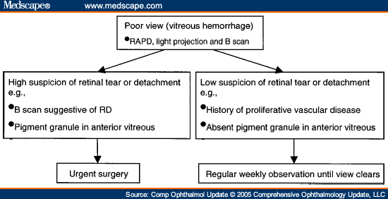
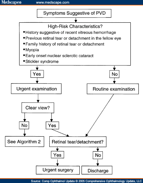
Diagnostic testing Posterior vitreous detachment is usually diagnosed with a dilated eye examination. However, if the vitreous gel is very clear, it may be hard to see the PVD without additional testing, such as optical coherence tomography (OCT) or ocular ultrasound (see Figure 2).
What is the meaning of vitreous detachment?
A vitreous detachment is a condition in which a part of the eye called the vitreous shrinks and separates from the retina. The vitreous is a gel-like substance that fills the inside of the eye ball. The retina is a light-sensitive area at the back of the eye.
When is PVD complete?
Acute PVD usually develops suddenly, becoming complete within weeks of onset of symptoms. A PVD is considered 'partial' when the vitreous jelly is still attached at the macula/optic nerve head and 'complete' once total separation of the jelly from the optic nerve head has occurred.Sep 23, 2016
What is the difference between retinal detachment and vitreous detachment?
The main difference between a vitreous detachment and retinal detachment is the damage done to the retina. On its own, PVD does not harm vision. As long as the fibers are merely pulling on the retina, the quality of your eyesight should not be affected.
What causes posterior vitreous detachment PVD?
What are causes of PVD? Age is the primary cause of PVD. As you age, it becomes harder for the vitreous to maintain its original shape. The vitreous gel shrinks and becomes more liquid-like, yet the cavity between your lens and retina remains the same size.Jan 28, 2019
Can high blood pressure cause posterior vitreous detachment?
Posterior vitreous detachment, often because it causes a retinal tear (see below). Retinal macroaneurysms - swollen blood vessels on the retina, usually related to high blood pressure, atherosclerosis and smoking.Jul 31, 2018
Can stress cause posterior vitreous detachment?
As with retinal detachment, stress on its own cannot cause a posterior vitreous detachment (PVD). A PVD is simply a normal process of aging in which the vitreous gel that fills the eye separates from the back of the eye.Oct 28, 2020
What are the symptoms of vitreous detachment?
If you see dark specks or flashes of light, it's possible you could have posterior vitreous detachment (PVD), an eye problem many people have as they age. As you get older, a gel inside your eye -- called vitreous gel -- can shrink. It can slowly detach (pull away) from your retina.Nov 9, 2020
How long does it take for posterior vitreous detachment to complete?
As long as you do not develop a retinal tear or retinal detachment, a PVD itself does not pose a threat to sight loss and the floaters and flashes slowly subside for a majority of patients within 3-6 months.Jan 22, 2019
Can posterior vitreous detachment be caused by trauma?
Blunt trauma to the eye or eye surgery may cause a PVD as well. Patients usually notice a PVD as a sudden onset of new floaters. Additionally, flashes of light indicating pulling upon the retina can be noted. Sometimes, a PVD can result in a retinal tear, which in turn could cause a retinal detachment.Nov 4, 2015
What is the most common cause of retinal detachment?
There are many causes of retinal detachment, but the most common causes are aging or an eye injury. There are 3 types of retinal detachment: rhegmatogenous, tractional, and exudative.
What is the ICd 10 code for vitreous degeneration?
H43.819 is a billable diagnosis code used to specify a medical diagnosis of vitreous degeneration, unspecified eye. The code H43.819 is valid during the fiscal year 2021 from October 01, 2020 through September 30, 2021 for the submission of HIPAA-covered transactions.#N#The ICD-10-CM code H43.819 might also be used to specify conditions or terms like degeneration of posterior vitreous body, posterior capsular rupture, posterior vitreous detachment, vitreous degeneration, vitreous detachment , vitreous liquefaction, etc.#N#Unspecified diagnosis codes like H43.819 are acceptable when clinical information is unknown or not available about a particular condition. Although a more specific code is preferable, unspecified codes should be used when such codes most accurately reflect what is known about a patient's condition. Specific diagnosis codes should not be used if not supported by the patient's medical record.
When to use H43.819?
Unspecified diagnosis codes like H43.819 are acceptable when clinical information is unknown or not available about a particular condition. Although a more specific code is preferable, unspecified codes should be used when such codes most accurately reflect what is known about a patient's condition. Specific diagnosis codes should not be used ...
Coding Notes for H43.81 Info for medical coders on how to properly use this ICD-10 code
Inclusion Terms are a list of concepts for which a specific code is used. The list of Inclusion Terms is useful for determining the correct code in some cases, but the list is not necessarily exhaustive.
ICD-10-CM Alphabetical Index References for 'H43.81 - Vitreous degeneration'
The ICD-10-CM Alphabetical Index links the below-listed medical terms to the ICD code H43.81. Click on any term below to browse the alphabetical index.
Popular Posts:
- 1. icd 10 code for non palpable pedal pulses
- 2. icd 10 code for solar dermatitis
- 3. icd 10 cm code for periumbilical pain
- 4. icd 10 code for right below knee amputation with prosthesis
- 5. icd 10 code for flat affect
- 6. icd 10 code for history of hairy cell leukemia
- 7. encounter for medicaments management icd 10 code
- 8. icd 10 cm code for disc extrusion at l5-s1
- 9. icd 10 code for chemo induced nausea and vomiting
- 10. icd-10 code for orthostasis