Full Answer
What is the ICD 10 code for pulmonary infiltrates?
Pulmonary infiltrates; Pulmonary nodules, multiple; Standard chest x-ray abnormal; Tomography - chest abnormal; ICD-10-CM R91.8 is grouped within Diagnostic Related Group(s) (MS-DRG v 38.0): 204 Respiratory signs and symptoms; Convert R91.8 to ICD-9-CM. Code History. 2016 (effective 10/1/2015): New code (first year of non-draft ICD-10-CM)
What is the ICD 10 code for abnormal lung field?
2018/2019 ICD-10-CM Diagnosis Code R91.8. Other nonspecific abnormal finding of lung field. 2016 2017 2018 2019 Billable/Specific Code. R91.8 is a billable/specific ICD-10-CM code that can be used to indicate a diagnosis for reimbursement purposes.
What is the ICD 10 code for pulmonary insufficiency following surgery?
pulmonary insufficiency following surgery ( ICD-10-CM Diagnosis Code J95.1. Acute pulmonary insufficiency following thoracic surgery 2016 2017 2018 2019 Billable/Specific Code. Type 2 Excludes Functional disturbances following cardiac surgery (I97.0, I97.1-) J95.1- ICD-10-CM Diagnosis Code J95.2.
What is the ICD 10 code for pulmonary atrophy?
Diagnosis Index entries containing back-references to J98.4: Adhesions, adhesive (postinfective) K66.0 ICD-10-CM Diagnosis Code K66.0 Atrophy, atrophic (of) lung J98.4 (senile) Calcification lung (active) (postinfectional) J98.4 Calculus, calculi, calculous lung J98.4 Cavitation of lung - see also Tuberculosis, pulmonary nontuberculous J98.4

What is the ICD-10-CM code for cavitary lesion of lung?
J98. 4 is a billable/specific ICD-10-CM code that can be used to indicate a diagnosis for reimbursement purposes. The 2022 edition of ICD-10-CM J98. 4 became effective on October 1, 2021.
What is cavitary lesion of lung?
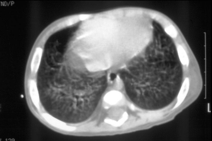
Right upper lobe cavitary lung lesion. A lung cavity is defined radiographically as a lucent area contained within a consolidation, mass, or nodule. 1. Cavities usually are accompanied by thick walls, greater than 4 mm.
What is a cavitary pulmonary nodule?
Cavitating nodular opacities in the course of rheumatic diseases are much rarer than interstitial pulmonary pneumonias and vasculitides. The nodules occur when epithelial cells cover a necrotic area, creating a necrobiotic nodule, which is the cause of the cavity.
What is the ICD-10 code for lung lesion?
ICD-10-CM Code for Solitary pulmonary nodule R91. 1.
What is the difference between pulmonary cavity and thoracic cavity?
The thoracic cavity is divided into two parts mediastinum and two pleural cavities. Pulmonary cavity is the subdivisional pleural lining on either side of the mediastinum and the space is occupied by the lungs.
What causes cavitary lesion?
Infections. Several groups of microorganisms may cause cavitary lesions: common bacteria (for example, Streptococcus p., Staph. aureus, Klebsiella p., H. influenzae); typical and atypical mycobacterium; fungi (for example, aspergillosis, pneumocystis j.); and parasites [9].
What is Cavitary disease?
Cavitary lung disease may result from several pathological processes, including suppurative necrosis, caseous necrosis, ischemic necrosis, displacement of lung tissue by cystic structures, and cystic dilatation of lung structures, vasculitis, or high-pressure traumas [1, 2].
What is the differential diagnosis of cavity in the lung?
During early radiology training, residents are introduced to the mnemonic “CAVITY” for the differential diagnosis of pulmonary cavitary lesions: cancer (bronchogenic carcinoma, especially squamous cell carcinoma), autoimmune (granulomatosis with polyangiitis or rheumatoid arthritis), vascular (pulmonary emboli – septic ...
What is the chest cavity?
The chest (thoracic or pleural) cavity is a space that is enclosed by the spine, ribs, and sternum (breast bone) and is separated from the abdomen by the diaphragm. The chest cavity contains the heart, the thoracic aorta, lungs and esophagus (swallowing passage) among other important organs.
What is Cavitary pneumonia?
Cavitary pneumonia is a rare complication of severe pneumonia in which normal lung tissue is replaced by a cavity. Most notably, it is associated with Mycobacterium tuberculosis.
What is diagnosis code R93 89?
ICD-10 code R93. 89 for Abnormal findings on diagnostic imaging of other specified body structures is a medical classification as listed by WHO under the range - Symptoms, signs and abnormal clinical and laboratory findings, not elsewhere classified .
What is the ICD-10 code for pulmonary mass?
For example, lung mass and multiple lung nodules are specifically indexed to code R91. 8, Other nonspecific abnormal finding of lung field.
Chronic pulmonary histoplasmosis capsulati. Nonrheumatic pulmonary valve insufficiency
Certain conditions have both an underlying etiology and hypothyoridism body system manifestations due to the underlying etiology.
Polyneuropathy in diseases classified elsewhere
Neoplasms CC14 Malignant neoplasms of lip, oral cavity Abnormal thyroid function study; Elevated thyroid stimulating hormone tsh ; Raised tsh level; Thyroid function tests abnormal. G62 Other and unspecified polyneuropathies.
Search Results
Chronic pulmonary histoplasma capsulatum; Chronic pulmonary histoplasmosis. Acute pulmonary histoplasmosis capsulati. I26 Pulmonary embolism I
Tuberculosis of lung
Congenital coarctation of pulmonary artery. Oral mouth lesion ; Oral lesion ; Oral mucosal lesion. Surfactant mutation of lung. Chronic pulmonary histoplasma capsulatum; Chronic pulmonary histoplasmosis. Blast injury of lung ; Blast lung.
Solitary pulmonary nodule
Search Results results found. Showing Pulmonary incompetence, non-rheumatic; Pulmonary valve regurgitation; Pulmonic valve regurgitation; Nonrheumatic pulmonary valve incompetence; Nonrheumatic pulmonary valve regurgitation.
Search Results
Pulmonary sarcoidosis; Sarcoidosis with lung involvement. Congenital absence of lung ; Congenital absence of lung lobe. Arteriovenous malformation, pulmonary ; Pulmonary arteriovenous malformation; Congenital pulmonary arteriovenous aneurysm. Complete lesion of conus medullaris. Search Results results found.
Secondary and unspecified malignant neoplasm of lymph nodes of head, face and neck
Pulmonary hypertension NOS. Kaposi sarcoma of bilateral lungs ; Kaposi sarcoma of left lung ; Kaposi sarcoma, bilateral lungs ; Kaposi sarcoma, left lung. Complete lesion of conus medullaris. Search Results results found. Acute pulmonary histoplasma capsulatum; Acute pulmonary histoplasmosis.

Popular Posts:
- 1. icd 10 code for removal of foreign body from ear
- 2. icd 10 code for kyphosis of thoracic region
- 3. icd 9 code for esophageal food bolus
- 4. icd 10 code for tpn status
- 5. icd code for abdominal bloating
- 6. icd 10 code for chronic osteomyelitis of foot
- 7. icd 10 code for osteoarthritis of left wrist
- 8. icd 9 code for acute respiratory infection
- 9. icd 10 code for status post intramedullary nailing of right femur fracture
- 10. icd 10 code for neck tightness