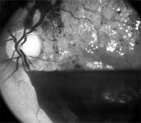
Retinal hemorrhage, left eye
H35. 62 is a billable/specific ICD-10-CM code that can be used to indicate a diagnosis for reimbursement purposes.What is the ICD-10 code for retinal hemorrhage?
ICD-10 | Retinal hemorrhage, unspecified eye (H35. 60)
What does retinal hemorrhage mean?
Retinal hemorrhage (UK English: retinal haemorrhage) is a disorder of the eye in which bleeding occurs in the retina, the light sensitive tissue, located on the back wall of the eye.
What is the ICD-10 code for subconjunctival hemorrhage?
31-33 Subconjunctival Hemorrhage. A subconjunctival hemorrhage is bleeding underneath the conjunctiva.Aug 5, 2016
What is macular hemorrhage?
Idiopathic Macular Hemorrhage is generally a disorder that primarily affecting patients younger than 40 years and can cause sudden unilateral loss of vision. It usually occurs in an otherwise healthy eye and mostly females. The exact pathogenesis of IMH remains unclear and poorly understood.
How is retinal hemorrhage diagnosed?
Fluorescein angiography may be used to take a picture of the inside of your eye. This test uses a dye that is injected into a vein in your hand or arm. The dye flows into the blood vessels of your retina, so your healthcare provider can see it clearly. Ultrasound may be used to show the bleeding inside your eye.
What is the difference between vitreous hemorrhage and retinal hemorrhage?
Decrease the gain to help differentiate between the two. Vitreous hemorrhage is usually less dense and will fade as the gain is decreased. It usually layers inferiorly with gravity. Ocular movements produce a rapid, staccato motion of the hemorrhage, unlike a retinal detachment that is stiffer and slower in movements.Dec 5, 2013
How does a subconjunctival hemorrhage heal?
Usually, treatment is unnecessary. A subconjunctival hemorrhage will resolve on its own within 7 to 14 days, gradually becoming lighter and less noticeable. Your doctor may recommend that you use artificial tears (Visine Tears, Refresh Tears, TheraTears) several times per day if your eye feels irritated.
What causes recurrent subconjunctival hemorrhage?
Certain medications or medical conditions can predispose an individual to recurrent subconjunctival hemorrhages. These conditions include diabetes, high blood pressure or hypertension, blood clotting disorders, and blood thinning medications like aspirin or Coumadin.
How long does a subconjunctival hemorrhage last?
Most people will not need any treatment. This condition often goes away on its own. Your subconjunctival hemorrhage will likely go away in a few weeks. It will first turn from red to brown, and then to yellow.
What is bilateral retinal hemorrhages?
Retinal hemorrhages in infancy are believed to be a cardinal sign of NAI. They may occur in up to 89% of infants with NAI. 1. They may result from direct head trauma or the acceleration and deceleration forces generated by the shaking of the head.
What causes retinal hemorrhaging?
Retinal hemorrhaging often occurs as a result of car accidents, sports accidents, falls from high locations, trip or slip and fall accidents, violent attacks, and similar traumatic events.Apr 13, 2017
What causes Submacular hemorrhage?
Submacular hemorrhage frequently results from a choroidal neovascular membrane secondary to age-related macular degeneration. Other conditions associated with CNVM, including myopia, trauma, ocular histoplasmosis and angioid streaks, can also lead to submacular hemorrhage.Jan 6, 2014
The ICD code H356 is used to code Retinal haemorrhage
Retinal hemorrhage is a disorder of the eye in which bleeding occurs into the light sensitive tissue on the back wall of the eye. A retinal hemorrhage can be caused by hypertension, retinal vein occlusion (a blockage of a retinal vein), or diabetes mellitus (which causes small fragile blood vessels to form, which are easily damaged).
Equivalent ICD-9 Code GENERAL EQUIVALENCE MAPPINGS (GEM)
This is the official approximate match mapping between ICD9 and ICD10, as provided by the General Equivalency mapping crosswalk. This means that while there is no exact mapping between this ICD10 code H35.62 and a single ICD9 code, 362.81 is an approximate match for comparison and conversion purposes.
What is the tissue that sends images to the brain?
The retina is a layer of tissue in the back of your eye that senses light and sends images to your brain. In the center of this nerve tissue is the macula. It provides the sharp, central vision needed for reading, driving and seeing fine detail.
What is the GEM crosswalk?
The General Equivalency Mapping (GEM) crosswalk indicates an approximate mapping between the ICD-10 code H35.62 its ICD-9 equivalent. The approximate mapping means there is not an exact match between the ICD-10 code and the ICD-9 code and the mapped code is not a precise representation of the original code.
Popular Posts:
- 1. icd 10 code for bilateral leg wounds
- 2. icd 10 code for gouty arthritis finger
- 3. icd 10 cm code for removal of uterus
- 4. icd 10 code for left rib series
- 5. icd-10-cm code for prothes
- 6. icd 10 code for hypercapnia
- 7. 2015 icd 10 code for intertrochanteric fracture right hip
- 8. icd 10 code for acute serositis
- 9. icd 10 code for fracture left femoral neck
- 10. is there an icd 10 code for normal hearing