Rheumatoid lung disease with rheumatoid arthritis of unspecified site. M05. 10 is a billable/specific ICD-10-CM code that can be used to indicate a diagnosis for reimbursement purposes.
What are the new ICD 10 codes?
The new codes are for describing the infusion of tixagevimab and cilgavimab monoclonal antibody (code XW023X7), and the infusion of other new technology monoclonal antibody (code XW023Y7).
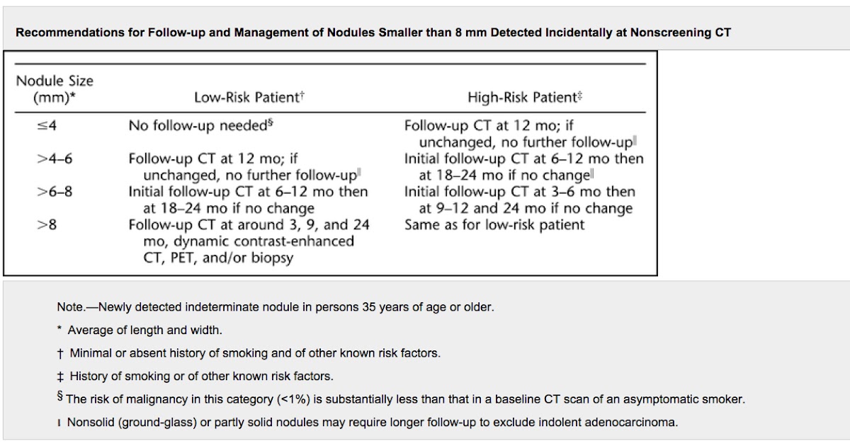
What is ICD 10 for pulmonary nodules?
- lung, solitary (subsegmental branch of the bronchial tree) R91.1
- pulmonary, solitary (subsegmental branch of the bronchial tree) R91.1
- solitary, lung (subsegmental branch of the bronchial tree) R91.1
What is the ICD 10 diagnosis code for?
The ICD-10-CM is a catalog of diagnosis codes used by medical professionals for medical coding and reporting in health care settings. The Centers for Medicare and Medicaid Services (CMS) maintain the catalog in the U.S. releasing yearly updates.
What is the diagnosis code for lung cancer?
- Acinar cell cystadenocarcinoma of lung
- Adenocarcinoma of lung
- Adenocarcinoma of lung, stage I
- Adenocarcinoma of lung, stage II
- Adenocarcinoma of lung, stage III
- Adenocarcinoma of lung, stage IV
- Adenosquamous cell carcinoma
- Anaplastic lymphoma kinase fusion oncogene negative non-small cell lung cancer
What is a rheumatoid nodule on the lung?
Lung nodules. Small lumps can form in the lungs (rheumatoid nodules), as well as in other parts of the body. Lung nodules usually cause no signs or symptoms, and they don't pose a risk of lung cancer. In some cases, however, a nodule can rupture and cause a collapsed lung.
What is the ICD-10 code for lung nodules?
ICD-10 code R91. 1 for Solitary pulmonary nodule is a medical classification as listed by WHO under the range - Symptoms, signs and abnormal clinical and laboratory findings, not elsewhere classified .
Are lung nodules common with RA?
RA-related lung complications are the most common extra-articular (“outside of the joints”) symptoms of RA and include pulmonary nodules (small growths in the lungs); pleural effusion (a buildup of fluid between the lung and chest wall); bronchiectasis (damage to the airways); and interstitial lung disease (ILD).
What is the ICD-10 code for multiple lung nodules?
For example, lung mass and multiple lung nodules are specifically indexed to code R91. 8, Other nonspecific abnormal finding of lung field.
What is the ICD 10 code for history of pulmonary nodules?
R91. 1 is a billable/specific ICD-10-CM code that can be used to indicate a diagnosis for reimbursement purposes. The 2022 edition of ICD-10-CM R91.
What is solitary pulmonary nodule in medical terms?
A solitary pulmonary nodule is a round or oval spot (lesion) in the lung that is seen with a chest x-ray or CT scan. This CT scan shows a single lesion (pulmonary nodule) in the right lung.
What lung disease is associated with rheumatoid arthritis?
Rheumatoid lung disease is a group of lung problems related to rheumatoid arthritis. The condition can include: Blockage of the small airways (bronchiolitis obliterans) Fluid in the chest (pleural effusions)
How big are rheumatoid nodules in lungs?
In the radiological semiology they are defined as rounded, multiple nodules, and more rarely as solitary nodules. They are preferentially located in the middle and superior peripheral lobe or pleural-based, with a size ranging from millimeters to 7cm.
Are rheumatoid lung nodules Spiculated?
Imaging of rheumatoid pulmonary nodules demonstrates nodules which are multiple in number with smooth borders, cavitation, satellite nodules and subpleural location with occasional subpleural rind. They are rarely spiculated in contrast to malignancy.
What is the ICD 10 code for right lower lobe lung mass?
ICD-10 Code for Malignant neoplasm of lower lobe, right bronchus or lung- C34. 31- Codify by AAPC.
What CPT code is R91 8?
Group 2CodeDescriptionR91.8Other nonspecific abnormal finding of lung field
What is the ICD 10 code for ASHD?
10 for Atherosclerotic heart disease of native coronary artery without angina pectoris is a medical classification as listed by WHO under the range - Diseases of the circulatory system .
Can a CT scan tell if a lung nodule is cancerous?
Can a CT scan tell if a lung nodule is cancerous? The short answer is no. A CT scan usually isn't enough to tell whether a lung nodule is a benign tumor or a cancerous lump. A biopsy is the only way to confirm a lung cancer diagnosis.
What is diagnosis code R91 8?
Other nonspecific abnormal finding of lung fieldICD-10 code R91. 8 for Other nonspecific abnormal finding of lung field is a medical classification as listed by WHO under the range - Symptoms, signs and abnormal clinical and laboratory findings, not elsewhere classified .
When is the ICd 10 code for rheumatoid nodule?
The 2021 edition of ICD-10-CM M06.3 became effective on October 1, 2020.
Where are subcutaneous nodules found?
Subcutaneous nodules seen in 20-30% of rheumatoid arthritis patients. They may arise anywhere on the body, but are most frequently found over the bony prominences .

Popular Posts:
- 1. icd 10 code for blood in cath bag
- 2. icd 10 code for cut with glass
- 3. icd 10 cm code for history of crohn disease and lupus,
- 4. icd 10 code for volvodynia
- 5. icd 10 code for s p severe brain injury
- 6. icd 10 code for carpal tunnel syndrome bilateral
- 7. icd 10 code for cecal polyp
- 8. icd 10 code for right shoulder ligament tear
- 9. 2017 icd 10 code for deviated nasal septum
- 10. icd code for tardive dyskinesia