The ICD-10-CM code M71. 38 might also be used to specify conditions or terms like arthropathy of lumbar facet joint, synovial cyst of lumbar spine or synovial cyst of sacrum.
What causes synovial cyst?
ICD-10-CM Diagnosis Code M71.2 Synovial cyst of popliteal space [Baker] synovial cyst of popliteal space with rupture (M66.0) ICD-10-CM Diagnosis Code M42.16 [convert to ICD-9-CM] Adult osteochondrosis of spine, lumbar region Adult osteochondrosis of lumbar spine ICD-10-CM Diagnosis Code M42.06 [convert to ICD-9-CM]
What is the recovery time for a synovial cyst surgery?
ICD-10-CM Diagnosis Code M71.38 [convert to ICD-9-CM] Other bursal cyst, other site. Synovial cyst of lumbar spine; Synovial cyst of sacrum; Synovial cyst, lumbar spine; Synovial cyst, sacrum. ICD-10-CM Diagnosis Code M71.38. Other bursal cyst, other site.
What is synovial cyst?
Apr 29, 2020 · Other bursal cyst, other site M71. 38 is a billable/specific ICD-10-CM code that can be used to indicate a diagnosis for reimbursement purposes. What is a lumbar synovial cyst? Synovial cysts are abnormal fluid-filled sacs in joints in the spine. These cysts are benign, which means they are not cancerous.
What is a synovial cyst of the lumbar spine?
Oct 01, 2021 · Synovial cyst of sacrum Synovial cyst, lumbar spine Synovial cyst, sacrum ICD-10-CM M71.38 is grouped within Diagnostic Related Group (s) (MS-DRG v39.0): 557 Tendonitis, myositis and bursitis with mcc 558 Tendonitis, myositis and bursitis without mcc Convert M71.38 to ICD-9-CM Code History
What is the ICD 10 code for synovial cysts?
Synovial cyst of popliteal space [Baker], unspecified knee M71. 20 is a billable/specific ICD-10-CM code that can be used to indicate a diagnosis for reimbursement purposes. The 2022 edition of ICD-10-CM M71. 20 became effective on October 1, 2021.
What is a synovial cyst in the spine?
Synovial cysts are abnormal fluid-filled sacs in joints in the spine. These cysts are benign, which means they are not cancerous. Synovial cysts typically develop as a result of degenerative changes that occur with aging. They can be found throughout the spine, but are most common in the lumbar region (low back).
How is a synovial cyst diagnosed?
Synovial Cyst Diagnosis The synovial cyst is best visualized on an MRI scan of the spine. It shows up as a hyperintense lesion that has the same signal intensity as water. X-rays, including flexion/extension motion x-rays, are also important to rule out any spinal instability.
What is the best way to treat a synovial cyst lumbar?
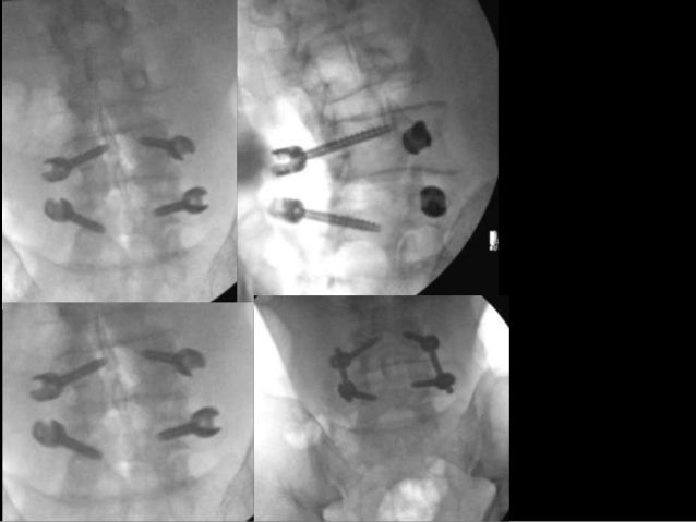
The most reliable treatment method for a synovial cyst is to remove the cyst and then fuse the joint. Fusing the joint stops all the motion at that level of the spine, and without any motion, the cyst should not regenerate. This is the most reliable treatment, but it is also an extensive surgery for the patient.
What is a lumbar facet cyst?
The cysts arise from the zygapophyseal joints of the lumbar spine and commonly demonstrate synovial herniation with mucinous degeneration of the facet joint capsule. Lumbar facet cysts are most common at the L4-L5 level and often are associated with spondylosis and degenerative spondylolisthesis.
What is a cyst at the base of the spine called?
There's a type of cyst you can get at the bottom of your tailbone, or coccyx. It's called a pilonidal cyst, and it can become infected and filled with pus. Once infected, the technical term is “pilonidal abscess,” and it can be painful.Mar 9, 2021
What causes lumbar cyst?
The cause of spinal cysts is unknown, but they may result from degeneration and instability of the spine in areas subjected to repetitive motion, particularly the joints in the lumbar region. Patients with spinal cysts may have other degenerative conditions of the spine, such as arthritis and disk disease.
How common are lumbar synovial cysts?
Bilateral lumbar facet cysts are rare, with reported frequency of 4% in surgically treated patients for lumbar facet cyst at the Mayo clinic over 24 years [5]. In the lumbar spine, synovial facet cysts are most commonly found at the L4–L5 level (68.4%) followed by the L5–S1 level (21.1%) [6, 7].
Can you feel a lumbar synovial cyst?
Many patients with synovial cysts experience no symptoms. Among those who do, common symptoms include: Lower back pain. Leg pain, also called sciatica, which can occur in one or both legs, and may radiate down the back of the leg and into the foot.
Can synovial cyst cause back pain?
Synovial cysts of the spine aren't deadly or cancerous and often don't produce symptoms. Symptoms that may occur include back pain or numbness, tingling, or cramping in the legs. There are treatments to help reduce discomfort, such as medication, activity modification, and injections.
Can a synovial cyst go away on its own?
Many synovial cysts will resolve on their own with conservative, non-operative treatments including rest, anti-inflammatory medications, or interventional pain management (steroidal injections). Occasionally, physical therapy may be recommended. Sometimes a physician will feel that draining the cyst is necessary.Apr 21, 2020
Do spinal synovial cysts go away?
Treatment options for spinal cysts These treatments are often effective at providing temporary relief, and in many cases, drainage of the cyst will cause it to go away on its own. Though uncommon, some cysts will not go away and will continue to cause problems such as pain, numbness or weakness.Apr 29, 2019
What is a synovial cyst?
A synovial cyst of the spine is a fluid-filled sac that develops along the spine. It’s the result of degeneration of a facet joint of the vertebrae of the spine. Most synovial cysts develop in the lumbar spine. M71.38, Other bursal cyst, other site.
What is the largest part of a vertebra?
Body: The body is the largest part of a vertebra, it is slightly cylindrical in shape, and faces interior of body. Its upper and lower surfaces are flattened and rough, and each presents a rim around its circumference. Foramen: A natural opening or passage, through a bone. Pedicles.
Where are upper and lower body parts located?
Where the general body part values “upper” and “lower” are provided as an option in the Upper Arteries, Lower Arteries, Upper Veins, Lower Veins, Muscles and Tendons body systems, “upper” or “lower “specifies body parts located above or below the diaphragm respectively. Example.
Where are the pedicles located?
: The pedicles are two short, thick processes, which project backward, one on either side, from the upper part of the body, at the junction of its posterior and lateral surfaces. The concavities above and below the pedicles are named the vertebral notches.

Popular Posts:
- 1. icd 10 code for driving safety
- 2. icd 10 code for abdominal pain possible appendix
- 3. icd-10-cm do you code for healed ulcer
- 4. icd 10 code for swelling left foot
- 5. icd 10 code for internal hemorrhoids unspecified
- 6. icd 10 code for fall with injury
- 7. icd-10 code for sudden death
- 8. 2019 icd 10 code for cpr
- 9. icd 10 code for bicuspid aortic valve
- 10. icd 10 code for dilated idiopathic