I77.810
What is a mild unfolding of the thoracic aorta?
What is a mild unfolding of the thoracic aorta? Aortic unfolding. Aortic unfolding is an abnormality visible on a chest X-ray, that shows widening of the mediastinum which may mimic the appearance of a thoracic aortic aneurysm. With aging, the ascending portion of the thoracic aorta increases in length by approximately 12% per decade, whereas the diameter increases by just 3% per decade.
What is new in dilatation of the ascending aorta?
- Causes of Dilatation of the Ascending Aorta. In patients with aortic dilatation, the aortic wall can be weakened by cystic media degeneration. ...
- Management and Follow-Up in Patients With Aortic Dilatation. ...
- Operative Treatment. ...
- Genetic Counseling. ...
- Conclusions. ...
- Follow-Up of Our Patient. ...
- Disclosures
- Footnotes. ...
What is thoracic aortic dilation?
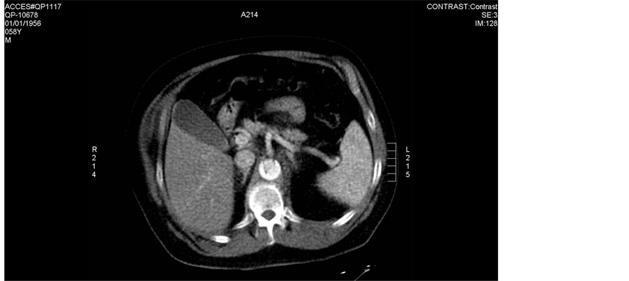
Thoracic aortic aneurysm (TAA) is a life-threatening condition that causes significant short- and long-term mortality due to rupture and dissection. For the thoracic aorta , a diameter greater than 3.5 cm is generally considered dilated, whereas greater than 4.5 cm would be considered aneurysmal.
What does unfolding of descending thoracic aorta mean?
Tortuosity of the descending thoracic aorta is a condition in which the aorta is misshapen and is characterized by abnormalities in blood vessels, particularly in arteries, says Genetics Home Reference. The descending thoracic aorta is one of four sections of the biggest heart artery called the aorta, notes WebMD.
What is the term for a bulge in the wall of an artery?
What causes a narrowing of the arteries?
About this website

What is the ICD-10 code for dilated aorta?
Q25.44Q25. 44 is a billable/specific ICD-10-CM code that can be used to indicate a diagnosis for reimbursement purposes.
What is thoracic aorta dilation?
Thoracic aortic aneurysm (TAA) is a life-threatening condition that causes significant short- and long-term mortality due to rupture and dissection. Aneurysm is defined as dilatation of the aorta of greater than 150% of its normal diameter for a given segment.
Is aortic dilation the same as aortic aneurysm?
Nevertheless, by common convention, aortic dilatation refers to a dimension that is greater than the 95th percentile for the normal person age, sex and body size. In contrast, an aneurysm is defined as a localized dilation of the aorta that is more than 50% of predicted (ratio of observed to expected diameter ≥ 1.5).
Is ascending aorta the same as thoracic aorta?
The entire aorta divides into two parts: the thoracic aorta and the abdominal aorta. The ascending aorta, along with the aortic arch and the descending aorta, makes up the thoracic aorta.
Where is the thoracic aorta?
The thoracic aorta runs from the aortic arch to the diaphragm, which is the point of separation between the chest cavity and the abdominal cavity. It provides blood to the muscles of the chest wall and the spinal cord.
What is mild aortic dilation?
A mild to moderately dilated ascending aorta was defined as having an aorta ascendens dimension between 40 mm to 45 mm on the computer tomography.
What is the ICD 10 code for thoracic aortic aneurysm?
ICD-10 code I71. 2 for Thoracic aortic aneurysm, without rupture is a medical classification as listed by WHO under the range - Diseases of the circulatory system .
What is aneurysmal dilatation of the ascending thoracic aorta?
An ascending aortic aneurysm is a weak spot in the top part of your aorta, which is the main artery in your body. The aneurysm bulges outward, and may cause your blood vessel wall to tear or break open. It's a life-threatening condition.
What is a thoracic aortic aneurysm?
A thoracic aortic aneurysm is a weakened area in the upper part of the body's main blood vessel (aorta). Aneurysms can develop anywhere in the aorta. A thoracic aortic aneurysm is a weakened area in the body's main artery (aorta) in the chest.
How common is dilated aorta?
66% of our patients were males and 34% females. 146 patients were found to have aortic dilatation. Therefore, the incidence of aortic dilatation was 6.8% in our study population.
What causes dilation of the ascending aorta?
Aneurysm formation can also be caused by chronic dissection, trauma, aortic surgery (false aneurysm), and cardiopulmonary resuscitation. Lastly, aortic dilatation can be caused by inflammatory diseases as bacterial or fungal aortitis, Takayasu arteritis, and giant cell arteritis.
What is a dilated aortic arch?
An aortic arch aneurysm is a bulge in the portion of the aorta closest to the heartlocated away from the heart and can involve the blood vessels that supply to your head and neck.
How serious is a dilated aorta?
Otherwise known as an aortic root aneurysm, a dilated aortic root is when the first section of the aorta, where the aortic valve resides, becomes enlarged. When this enlargement reaches a critical size, there is a risk of it rupturing or tearing, leading to a life-threatening situation.
What is the treatment for a dilated aorta?
The most common type of surgery is open abdominal or chest repair, where the doctor opens up your chest or abdomen, depending on where the problem is, removes the bulge in your aorta, and replaces it with a fabric tube called a graft.
What are the symptoms of a dilated aorta?
However as it enlarges, aortic dilation symptoms like these may include:Pain in the chest or upper back. The pain may be deep, aching, gnawing, and/or throbbing, and may last for hours or days. ... Shortness of breath, a raspy voice.Pain in the left shoulder or between the shoulder blades.Pain in the groin.
How common is a dilated aorta?
66% of our patients were males and 34% females. 146 patients were found to have aortic dilatation. Therefore, the incidence of aortic dilatation was 6.8% in our study population.
2022 ICD-10-CM Diagnosis Code I77.810: Thoracic aortic ectasia
Free, official coding info for 2022 ICD-10-CM I77.810 - includes detailed rules, notes, synonyms, ICD-9-CM conversion, index and annotation crosswalks, DRG grouping and more.
2022 ICD-10-CM Diagnosis Code I77.81: Aortic ectasia
A type 1 excludes note is a pure excludes. It means "not coded here". A type 1 excludes note indicates that the code excluded should never be used at the same time as I77.81.A type 1 excludes note is for used for when two conditions cannot occur together, such as a congenital form versus an acquired form of the same condition.
I77.810 - ICD-10 Code for Thoracic aortic ectasia - Billable
I77.810 is a valid billable ICD-10 diagnosis code for Thoracic aortic ectasia.It is found in the 2022 version of the ICD-10 Clinical Modification (CM) and can be used in all HIPAA-covered transactions from Oct 01, 2021 - Sep 30, 2022.. ↓ See below for any exclusions, inclusions or special notations
ICD-10-CM Code I77.810 - Thoracic aortic ectasia
This is the official exact match mapping between ICD9 and ICD10, as provided by the General Equivalency mapping crosswalk. This means that in all cases where the ICD9 code 447.71 was previously used, I77.810 is the appropriate modern ICD10 code.
2022 ICD-10-CM Code I71.2 - Thoracic aortic aneurysm, without rupture
I71.2 is a billable diagnosis code used to specify a medical diagnosis of thoracic aortic aneurysm, without rupture. The code I71.2 is valid during the fiscal year 2022 from October 01, 2021 through September 30, 2022 for the submission of HIPAA-covered transactions.
What is the term for a bulge in the wall of an artery?
Aneurysm - a bulge or "ballooning" in the wall of an artery. Atherosclerosis - a disease in which plaque builds up inside your arteries. Plaque is made up of fat, cholesterol, calcium, and other substances found in the blood. Blood clots, including deep vein thrombosis and pulmonary embolism.
What causes a narrowing of the arteries?
Coronary artery disease and carotid artery disease , diseases that involve the narrowing or blockage of an artery. The cause is usually a buildup of plaque. Raynaud's disease - a disorder that causes the blood vessels to narrow when you are cold or feeling stressed.
Popular Posts:
- 1. icd 10 code for crush injury distal left thumb
- 2. icd 10 code for e11.42
- 3. icd 10 code for bcc of nose
- 4. icd 10 code for no free fluid.
- 5. icd 10 code for fungal infection in toe web spaces
- 6. icd 10 code for r wrist injury
- 7. icd-9-cm code for situs inversus abdominalis
- 8. icd 10 code for contusion right middle finger
- 9. icd 10 code for presence of port
- 10. icd 10 code for pulmonary fibrosis exacerbation