Vitreomacular adhesion, left eye. H43.822 is a billable/specific ICD-10-CM code that can be used to indicate a diagnosis for reimbursement purposes. The 2019 edition of ICD-10-CM H43.822 became effective on October 1, 2018.
ICD-10: H43. 821 Vitreomacular adhesion, right eye.Apr 19, 2022
Full
AnswerWhat is the ICD 10 code for vitreomacular adhesion?
· The 2022 edition of ICD-10-CM H43.82 became effective on October 1, 2021. This is the American ICD-10-CM version of H43.82 - other international versions of ICD-10 H43.82 may differ. Applicable To Vitreomacular traction The following code (s) above H43.82 contain annotation back-references that may be applicable to H43.82 : H00-H59
What is vitreomacular traction?
· Vitreomacular adhesion, left eye 2016 2017 2018 2019 2020 2021 2022 Billable/Specific Code Adult Dx (15-124 years) H43.822 is a billable/specific ICD-10-CM code that can be used to indicate a diagnosis for reimbursement purposes. The 2022 edition of ICD-10-CM H43.822 became effective on October 1, 2021.
What is the ICD 10 code for posterior vitreous detachment?
Vitreomacular adhesion. ICD-10-CM H43.82. https://icd10coded.com/cm/H43.82/. Includes: Vitreomacular traction.
What is the ICD 10 code for vitreous abscess?
· Vitreomacular adhesion, unspecified eye. 2016 2017 2018 2019 2020 2021 2022 Billable/Specific Code Adult Dx (15-124 years) H43.829 is a billable/specific ICD-10-CM code that can be used to indicate a diagnosis for reimbursement purposes. The 2022 edition of ICD-10-CM H43.829 became effective on October 1, 2021.

What is VMT of the eye?
Vitreomacular traction (VMT) happens when vitreous has an unusually strong attachment to the retina in the back of the eye. As the eye ages, the vitreous doesn't detach completely from the macula as it should. The vitreous then pulls on the macula, which can damage the macula and threaten vision.
What is the ICD-10 code for vitrectomy?
Filtering (vitreous) bleb after glaucoma surgery status The 2022 edition of ICD-10-CM Z98. 83 became effective on October 1, 2021. This is the American ICD-10-CM version of Z98.
What is the ICD-10 code for vitreous detachment?
CASE 2 – POSTERIOR VITREOUS DETACHMENT (PVD) What ICD-10 code(s) should be used There are two valid diagnoses: H43. 811 (Vitreous degeneration, right eye) and Z96. 1 (Presence of intraocular lens; pseudophakia).
How is vitreomacular traction treated?
Some cases of VMT may spontaneously resolve. For patients whose symptoms are severe enough to require intervention, pars plana vitrectomy surgery is one treatment option. The procedure involves the manual release of vitreous attachment and alleviation of traction, but it is invasive and inconvenient to most patients.
What is the ICD-10 code for traction retinal detachment?
H33.40Traction detachment of retina, unspecified eye H33. 40 is a billable/specific ICD-10-CM code that can be used to indicate a diagnosis for reimbursement purposes. The 2022 edition of ICD-10-CM H33. 40 became effective on October 1, 2021.
What is the CPT code for vitrectomy?
If vitrectomy is performed with the removal of the internal limiting membrane for the repair of a MH, the CPT code that should be used is 67042—vitrectomy, mechanical, pars plana approach; with removal of internal limiting membrane of retina (eg, for repair of MH, diabetic macular edema), includes, if performed, ...
What is the ICD-10 code for PVD?
ICD-10 | Peripheral vascular disease, unspecified (I73. 9)
What happens when the vitreous separates from the retina?
When your vitreous detaches, strands of the vitreous often cast new shadows on your retina — and those shadows appear as floaters. You may also notice flashes of light in your side (peripheral) vision. Sometimes, vitreous detachment causes more serious eye problems that need treatment right away.
What causes posterior vitreous detachment?
Posterior vitreous detachment (PVD) occurs when the gel that fills the eyeball separates from the retina. It's a natural, normal part of aging. PVD can cause floaters or flashes in your sight, which usually become less noticeable over time. The condition isn't painful, and it doesn't cause vision loss on its own.
Is vitreomacular traction the same as vitreomacular adhesion?
It sticks -- or adheres -- to the macula. This is vitreomacular adhesion. It can hold on so strongly, it pulls on the macula (your eye doctor may call this “traction”). When that happens, it can affect vision -- what doctors call symptomatic VMA.
How common is VMT?
VMT only occurs in about 1 in 4400 people. The occurrence of VMT in patients with diabetic retinopathy, age-related macular degeneration, and other macular diseases is much higher. It occurs in women slightly more often than men and can happen at any age, in any race.
Is vitreomacular traction an emergency?
Treatment is not necessary if symptoms are mild. However, when vitreomacular traction causes visual loss or the symptoms start to interfere with normal daily activities, then surgery is recommended. In most cases, vitrectomy surgery is the most effective treatment to release the vitreomacular traction.
What causes vitreomacular traction syndrome?
As we age, the vitreous becomes more watery. It may eventually separate from the retina in a process known as posterior vitreous detachment. If the vitreous doesn't fully separate from the retina, the remaining partially attached vitreous can pull and distort the macula. This is vitreomacular traction.
How long is vitrectomy recovery?
After the surgery, your eye may be swollen, red, or tender for several weeks. You might have some pain in your eye and your vision may be blurry for a few days after the surgery. You will need 2 to 4 weeks to recover before you can do your normal activities again.
What causes macular traction?
As we grow older the thick vitreous gel that fills the eye shrinks and pulls away from the macula. Sometimes as the vitreous pulls away from the macula, it remains partially stuck and pulls on the surface. This pulling and distortion of the normal macular structure is called vitreomacular traction (VMT).
Why is vitrectomy performed?
Vitrectomy is a surgical procedure undertaken by a specialist where the vitreous humor gel that fills the eye cavity is removed to provide better access to the retina. This allows for a variety of repairs, including the removal of scar tissue, laser repair of retinal detachments and treatment of macular holes.
What is the ERM in vitreomacular traction?
An epiretinal membrane (ERM) is commonly associated with both the vitreofoveal and vitreomacular traction. It has been shown to proliferate from the retinal surface, coursing up the cone of attached vitreous, and then growing along the back surface of the detached perifoveal hyaloid. ERM is thought to exacerbate VMT by.
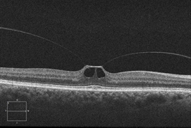
What is OCT in vitreomacular interface?
Optical coherence tomography (OCT) allows noninvasive visualization and imaging of vitreomacular interface and is an important tool in the diagnosis and management of VMT syndrome, especially with the advent of non-surgical management with pharmacologic vitreolysis. The OCT in VMT demonstrates a partial posterior vitreous detachment with persistent vitreous attachment to the fovea alone or in addition to the macula and/or the optic disc margin. The posterior hyaloid usually appears as a hyperreflective and thickened line/band on OCT located posterior to the hyporeflective vitreous and anterior to the retina. The appearance of VMT is accentuated when the sensitivity if the image is increased (by making the image lighter in the OCT machine). It courses in a conical pattern posteriorly to attach to the macula. According to Chang et al., the hyperreflective band corresponds to the bridging fibrocellular proliferative tissue.
What is the term for the adherence of the posterior hyaloid to the retina?
The anomalous adherence of the posterior hyaloid to the retina can be either a primary abnormality or develop secondary to cellular proliferation from cortical vitreous remnants after a partial posterior vitreous detachment, also known as vitreoschisis, or from concurrent diseases (e.g. proliferative diabetic retinopathy) that provide a fibrovascular scaffold for cellular proliferation and contraction. An epiretinal membrane (ERM) is commonly associated with both the vitreofoveal and vitreomacular traction. It has been shown to proliferate from the retinal surface, coursing up the cone of attached vitreous, and then growing along the back surface of the detached perifoveal hyaloid.
What is the diameter of the macula?
certain areas of the macula – mainly to the foveola (500 μm diameter area in the center of macula) and the margin of the fovea (circumference of the 1500 μm diameter fovea).
Which part of the retina is most firmly attached to the vitreous?
The vitreous is most firmly attached to the retina in areas where the ILM is the thinnest including
When anterior vitreous pull and weakening of attachments occur synchronously, a normal posterior vitre
When anterior vitreous pull and weakening of attachments occur synchronously, a normal posterior vitreous detachment (PVD) occurs. However, when these occur asynchronously (tractional component preceding or proceeding faster than the vitreoretinal detachment), an anomalous PVD develops which can result in VMT and other vitreoretinal diseases. As noted in the table above, the VMT progression can correlate with various stages of FTMH.
What is VFT in a vitreomacular?
Many authors have identified a subset of VMT called vitreofoveal traction (VFT) where the vitreomacular attachment is limited to a focal foveal region (as shown in the figure above) differentiating it from classic VMT. Prior classifications of VMT are summarized in the section on OCT presented above.
What causes VMT in the eye?
VMT is usually caused by part of the vitreous remaining stuck to the macula during a posterior vitreous detachment. In healthy eyes, VMT is not common. People with certain eye diseases may be at a higher risk for VMT, including those with: high myopia (extreme nearsightedness)
What is the term for the breakdown of tissues in the back of the eye?
high myopia (extreme nearsightedness) age-related macular de generation (AMD) (a breakdown of tissues in the back of the eye) diabetic eye disease (disease that affects the blood vessels in the back of the eye) retinal vein occlusion (a blockage of veins in the retina)
What is it called when the retina separates from the eye?
The vitreous, over time, separates completely from the retina. This is called a posterior vitreous detachment (PVD) and is usually a normal part of aging. It happens to most people by age 70. In some people with PVD, the vitreous doesn’t detach ...
Popular Posts:
- 1. icd 10 code for acute bronchospasm with asthma
- 2. icd 10 cm code for disorder of soft tissue
- 3. icd code for chronic ischemic heart disease
- 4. icd 10 code for laceration right little finger
- 5. icd 10 code for newborn reflux
- 6. icd 10 code for cirrhosis of family history of diabetes
- 7. icd 10 code for small finger fracture
- 8. icd 10 cm code for burning in esophagus
- 9. what is the icd 10 code for poor compliance with diet and medication
- 10. icd 10 code for chronic leg pain