Age-related macular degeneration ICD-9-CM diagnosis codes nn362.50 (macular degeneration [senile], unspecified) n362.51 (nonexudative senile macular degeneration)
Types of Macular Degeneration
Macular degeneration generally starts in the dry form and is classified to ICD-9-CM code 362.51.Mar 5, 2007How do you cure macular degeneration?
Other dietary steps to take include:
- Avoid beta carotene.
- Eat more vegetables, especially leafy greens.
- Reduce sugar intake significantly.
- Consume more omega-3 fatty acid foods, like fish.
- Eat more fruit, especially high-fiber fruit.
What are common causes of macular degeneration?
Wet macular degeneration
- Overview. As macular degeneration develops, clear, normal vision (shown left) becomes impaired by a general haziness.
- Symptoms. Wet macular degeneration symptoms usually appear suddenly and worsen rapidly. ...
- Causes. ...
- Risk factors. ...
- Complications. ...
- Prevention. ...
How to code for macular degeneration?
- Quit smoking — or don’t start
- Get regular physical activity
- Maintain a healthy blood pressure and cholesterol levels
- Eat healthy foods, including leafy green vegetables and fish
How to manage your macular degeneration?
Managing and Monitoring Dry AMD
- See your eye doctor regularly. Khurana says he typically sees people with dry AMD every six months. ...
- Monitor your vision at home for any changes. Your eye doctor may suggest that you use an Amsler grid, which looks like a piece of graph paper. ...
- Stop smoking. If you smoke, one of the many reasons to quit is that it can worsen AMD. ...
- Take eye vitamins. ...

What is the ICD code for macular degeneration?
ICD-10-CM Code for Exudative age-related macular degeneration H35. 32.
What does Nonexudative age-related macular degeneration mean?
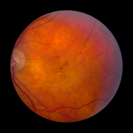
Nonexudative AMD is characterized by the degeneration of the retina and the choroid in the posterior pole due to either atrophy or RPE detachment. The atrophy is generally preceded (or coincident in some cases) by the presence of yellow extracellular deposits adjacent to the basal surface of the RPE called drusen.
What is the ICD-10 code for dry macular degeneration?
3131: Nonexudative age-related macular degeneration, bilateral, early dry stage.
What is the ICD 9 code for retinal detachment?
361.9Short description: Retinal detachment NOS. ICD-9-CM 361.9 is a billable medical code that can be used to indicate a diagnosis on a reimbursement claim, however, 361.9 should only be used for claims with a date of service on or before September 30, 2015.
What are the stages of age-related macular degeneration?
Most people with AMD have dry AMD (also called atrophic AMD). This is when the macula gets thinner with age. Dry AMD happens in 3 stages: early, intermediate, and late.
What is the difference between wet and dry macular degeneration?
The main difference between wet vs dry macular degeneration is simple: dry macular degeneration is the more common type of eye disease and does less damage to your vision while wet macular degeneration can result in serious vision loss.
What is the ICD-10-CM code for osteopenia?
Under ICD-10-CM, the term “Osteopenia” is indexed to ICD-10-CM subcategory M85. 8- Other specified disorders of bone density and structure, within the ICD-10-CM Alphabetic Index.
What is the ICD-10 code for dry eyes?
ICD-10-CM Code for Dry eye syndrome H04. 12.
What is the ICD-10 code for ASHD?
ICD-10 Code for Atherosclerotic heart disease of native coronary artery without angina pectoris- I25. 10- Codify by AAPC.
What is macula off retinal detachment ICD-10?
Retinal detachment with giant retinal tear, left eye H33. 032 is a billable/specific ICD-10-CM code that can be used to indicate a diagnosis for reimbursement purposes. The 2022 edition of ICD-10-CM H33. 032 became effective on October 1, 2021.
What is macula off retinal detachment?
Macular detachment was defined as detachment involving the fovea with any resulting loss of central Snellen visual acuity. The series was an unbiased selection of cases under the care of two consultants (THW: n=110 and DAHL: n=75) in a vitreoretinal unit of a teaching hospital.
Which of the following diagnoses is reported with code H27 00?
2022 ICD-10-CM Diagnosis Code H27. 00: Aphakia, unspecified eye.
Coding For Laterality in AMD
When you use the codes for dry AMD (H35.31xx) and wet AMD (H35.32xx), you must use the sixth character to indicate laterality as follows:1 for the...
Coding For Staging in Dry AMD
The codes for dry AMD—H35.31xx—use the seventh character to indicate staging as follows:H35.31x1 for early dry AMD—a combination of multiple small...
Defining Geographic Atrophy
When is the retina considered atrophic? The Academy Preferred Practice Pattern1 defines GA as follows:The phenotype of central geographic atrophy,...
Coding For Geographic Atrophy
The Academy recommends that when coding, you indicate whether the GA involves the center of the fovea: Code H35.31x4 if it does and H35.31x3 if it...
Coding For Staging in Wet AMD
The codes for wet AMD—H35.32xx—use the sixth character to indicate laterality and the seventh character to indicate staging as follows:H35.32x1 for...
Coding for Laterality in AMD
When you use the codes for dry AMD (H35.31xx) and wet AMD (H35.32xx), you must use the sixth character to indicate laterality as follows:
Coding for Staging in Dry AMD
The codes for dry AMD—H35.31xx—use the seventh character to indicate staging as follows:
Defining Geographic Atrophy
When is the retina considered atrophic? The Academy Preferred Practice Pattern1 defines GA as follows:
Coding for Geographic Atrophy
The Academy recommends that when coding, you indicate whether the GA involves the center of the fovea: Code H35.31x4 if it does and H35.31x3 if it doesn’t, with “x” indicating laterality.
Coding for Staging in Wet AMD
The codes for wet AMD—H35.32xx—use the sixth character to indicate laterality and the seventh character to indicate staging as follows:
Focus on Payment Policy at AAO 2017
Introduction to Physician Payment Policy (Sym12). A panel will explain how new CPT codes are created and valued; how existing codes are targeted for reevaluation; the impact of new technology on the valuation of existing procedures; and the difference between CMS and commercial carrier coverage policies. When: Sunday, Nov. 12, 11:15 a.m.-12:15 p.m.
How to tell if you have wet macular degeneration?
The symptoms for wet macular degeneration include the following: • visual distortions; • decrease in or loss of central vision; and. • central blurry spot. Diagnosis. Macular degeneration is diagnosed during a thorough eye examination. The physician will look for drusen and mottled pigmentation in the macula.
What does a doctor look for in a macular test?
The physician will look for drusen and mottled pigmentation in the macula. In addition, the physician may have the patient look at an Amsler grid , which looks like a checkerboard. This test can determine the location and extent of the macular damage.
What is the cause of vision loss in older people?
Macular degeneration is a chronic eye condition that occurs because of deterioration of the tissue in the macula—the central portion of the retina responsible for central vision. Sometimes referred to as age-related macular degeneration , it is the leading cause of severe vision loss in people aged 60 and older.
What test is used to determine the extent of damage to the macula?
The physician may also choose to perform a fluorescein angiography to evaluate the extent of the damage to the macula. Another test that may be done is optical coherence tomography which identifies the area of retinal thickening or thinning. It is extremely beneficial to diagnosis macular degeneration early.
Can macular degeneration cause blindness?
Although it does not lead to total blindness, it can cause blurred central vision or a blind spot in the center of the visual field. Typically, the condition does not affect peripheral vision. Macular degeneration is a gradually progressive disease that is nonreversible.
Can macular degeneration be reversed?
Although the damage from macular degeneration cannot be reversed, early treatment can slow progression.
Popular Posts:
- 1. code for rash following measles vaccination icd 10
- 2. icd-10-cm pcs code for piv in cephalic vein ??
- 3. icd 10 cm code for for . prednisone
- 4. icd-10 code for atherosclerosis of coronary artery bypass
- 5. icd 10 code for post surgical ptca stent
- 6. icd 10 code for appy
- 7. icd 10 code for epilepsy, visual type
- 8. icd 10 code for atrial fibrillation paroxysmal
- 9. icd-9-cm code for depression with mood behavior
- 10. icd 10 code for involuntary commitment