D18.00 — Hemangioma
Hemangioma
A non-cancerous reddish tumor formed of excess blood vessels.
ICD-9-CM Diagnosis Code 228.00 : Hemangioma of unspecified site.
Full
AnswerWhat is the CPT code for Cherry angioma?
#1 When indicating a diagnosis code for cherry angiomas, should code D18.00 or code D18.01 be used. Not sure even though these are on the skin. Does anyone know the proper code to use.
What are cherry angiomas and how common are they?
Cherry angiomas are small, red bumps on your skin that are harmless to your overall health. Angiomas commonly appear after age 30 and can be removed if you don’t like how they look. What is a cherry angioma?
What are the treatment options for a Cherry angioma?
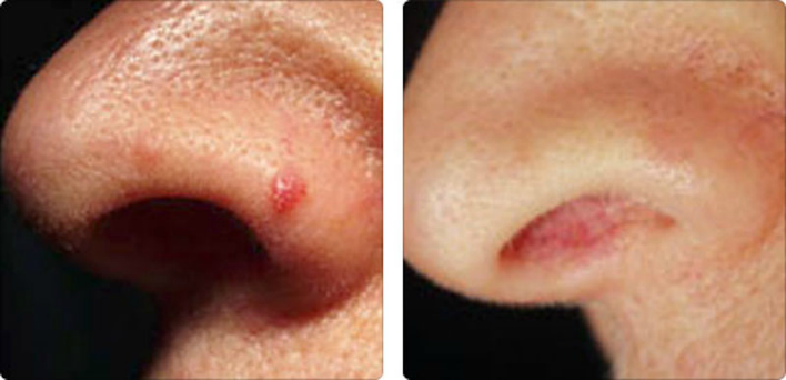
Cherry angiomas don’t typically need to be treated. However, if they're bothering you or bleeding frequently, they can be treated in non-invasive ways that cause minimal to no scarring. These treatments are also relatively painless. Electrodesiccation: The spot is touched with an electric needle that destroys the blood vessels.
What is the ICD-10 code for cherry angiomas?
D18.01When indicating a diagnosis code for cherry angiomas, should code D18. 00 or code D18. 01 be used.
What is the ICD-10 code for Angioma?
D18.01Hemangioma of skin and subcutaneous tissue D18. 01 is a billable/specific ICD-10-CM code that can be used to indicate a diagnosis for reimbursement purposes. The 2022 edition of ICD-10-CM D18. 01 became effective on October 1, 2021.
What ICD-9 codes?
ICD-9-CM is the official system of assigning codes to diagnoses and procedures associated with hospital utilization in the United States. The ICD-9 was used to code and classify mortality data from death certificates until 1999, when use of ICD-10 for mortality coding started.
What is ICD-9 code lesion of skin?
86.3 Other local excision or destruction of lesion or tissue of skin and subcuta - ICD-9-CM Vol.
Is Angioma the same as hemangioma?
Angioma or haemangioma (American spelling 'hemangioma') describes a benign vascular skin lesion. An angioma is due to proliferating endothelial cells; these are the cells that line the inside of a blood vessel.
What is an Angioma?
Angiomas are benign growths made up of small blood vessels. They can appear anywhere on the body. The three most common types are cherry angiomas, spider angiomas, and angiokeratomas.
Are ICD-9 codes still used in 2021?
CMS will continue to maintain the ICD-9 code website with the posted files. These are the codes providers (physicians, hospitals, etc.) and suppliers must use when submitting claims to Medicare for payment.
What is ICD-9 and ICD-10 difference?
ICD-9 uses mostly numeric codes with only occasional E and V alphanumeric codes. Plus, only three-, four- and five-digit codes are valid. ICD-10 uses entirely alphanumeric codes and has valid codes of up to seven digits.
What are ICD-9 10 and CPT codes?
ICD-10-CM diagnosis codes provide the reason for seeking health care; ICD-10-PCS procedure codes tell what inpatient treatment and services the patient got; CPT (HCPCS Level I) codes describe outpatient services and procedures; and providers generally use HCPCS (Level II) codes for equipment, drugs, and supplies for ...
What is the ICD-10 code for keratosis pilaris?
Acquired keratosis [keratoderma] palmaris et plantaris L85. 1 is a billable/specific ICD-10-CM code that can be used to indicate a diagnosis for reimbursement purposes. The 2022 edition of ICD-10-CM L85. 1 became effective on October 1, 2021.
What is the ICD-9 code for cellulitis?
ICD-9 code 682.9 for Cellulitis and abscess of unspecified sites is a medical classification as listed by WHO under the range -INFECTIONS OF SKIN AND SUBCUTANEOUS TISSUE (680-686).
What is the ICD-9 code for hemorrhoids?
ICD-9 Code 455.6 -Unspecified hemorrhoids without complication- Codify by AAPC.
Not Valid for Submission
228.09 is a legacy non-billable code used to specify a medical diagnosis of hemangioma of other sites. This code was replaced on September 30, 2015 by its ICD-10 equivalent.
ICD-9 Footnotes
General Equivalence Map Definitions The ICD-9 and ICD-10 GEMs are used to facilitate linking between the diagnosis codes in ICD-9-CM and the new ICD-10-CM code set. The GEMs are the raw material from which providers, health information vendors and payers can derive specific applied mappings to meet their needs.
How to diagnose cherry angioma?
Diagnosis. Doctors often diagnose a cherry angioma just by looking at it. They may request a biopsy if they suspect it's a potentially harmful skin growth instead. Your doctor may also want to check for a separate type of skin growth called spider angiomas.
What is a cherry angioma?
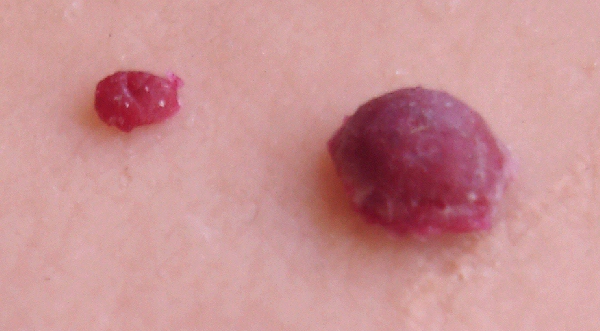
A cherry angioma is classified as a bright, cherry-red or purple spot, which is due to the dilated capillaries they’re made up of. They can range widely in size, from a tiny dot to several millimeters in diameter. Cherry angiomas are commonly round to oval-shaped.
How long does it take for a cherry angioma to grow back?
With any treatment procedure, it’s possible that a cherry angioma will grow back over time. If so, you can get it removed again.
Why do cherry angiomas appear in clusters?
Exposure to certain chemicals and gases in the environment can also cause cherry angiomas to appear in clusters. They're also more common in certain climates. 2. It’s also possible that hormones play a role in the appearance of cherry angiomas, as they're often discovered after childbirth.
How many cherry angiomas are there in a 70 year old?
But almost every 70-year-old has one or more of them , making these skin growths a frequent issue many people deal with. 1
What does it mean when you have spider angioma?
Most common during pregnancy and in children, spider angiomas that appear suddenly and in groups may be a warning sign for liver damage. 4 If your doctor isn't sure which type of angioma you have, they may run blood or imaging tests to check your liver health. Healthy Skin in Your 20s, 30s, 40s, and Beyond.
How to remove angioma from blood vessels?
Common removal methods include: 5. Electrodesiccation: The spot is touched with an electric needle that destroys and breaks up the blood vessels. Liquid nitrogen or cryotherapy : Using a probe, cold gas is sprayed on the angioma, causing it to fall off in a few hours.
What is a cherry hemangioma?
Cherry hemangiomas (also known as cherry angiomas, Campbell de Morgan spots, and senile hemangiomas) are the most common type of acquired benign vascular proliferation and are composed of thin-walled, dilated capillaries.
Do cherry hemangiomas require treatment?
Cherry hemangiomas are benign and thus do not require treatment unless irritated or bleeding (usually secondary to trauma), but are often of cosmetic concern to patients. The etiology of cherry hemangiomas is still poorly understood, with evidence supporting either a reactive or a neoplastic underpinning.
Is cherry hemangioma a risk factor?
The strongest risk factor for cherry hemangiomas is age. However, studies have also identified associations between cherry hemangiomas and toxic exposures including mustard gas and bromide, and with a variety of more severe health conditions including melanoma, nonmelanoma skin cancer, noncutaneous malignancy, immunosuppression, and dyslipidemia.
What is a benign skin lesion?
The majority of cases are congenital. A benign skin lesion consisting of dense, usually elevated masses of dilated blood vessels. A benign tumor of the blood vessels that appears on skin. A benign vascular neoplasm characterized by the formation of capillary-sized or cavernous vascular channels.
Is morphology included in the category and codes?
In a few cases, such as for malignant melanoma and certain neuroendocrine tumors, the morphology (histologic type) is included in the category and codes. Primary malignant neoplasms overlapping site boundaries.
Popular Posts:
- 1. icd 10 code for adductor policis tendionitit\s
- 2. icd 10 cm code for s/p vps
- 3. icd 10 code for nitroglycerin
- 4. icd 10 code for left calcaneal pain
- 5. icd 10 code for major traumatic injury
- 6. icd 10 medical code for subdural hematoma(nontraumatic subdural hemorrhage unspecified)
- 7. icd 10 code for laceration to right knee
- 8. icd 10 code for rt hand swelling
- 9. icd 10 code for verruca
- 10. icd 10 code for fibromuscular dysplasia