Full Answer
What are the new ICD 10 codes?
The new codes are for describing the infusion of tixagevimab and cilgavimab monoclonal antibody (code XW023X7), and the infusion of other new technology monoclonal antibody (code XW023Y7).
What are ICD 10 codes?
Why ICD-10 codes are important
- The ICD-10 code system offers accurate and up-to-date procedure codes to improve health care cost and ensure fair reimbursement policies. ...
- ICD-10-CM has been adopted internationally to facilitate implementation of quality health care as well as its comparison on a global scale.
- Compared to the previous version (i.e. ...
What is the ICD 10 diagnosis code for?
The ICD-10-CM is a catalog of diagnosis codes used by medical professionals for medical coding and reporting in health care settings. The Centers for Medicare and Medicaid Services (CMS) maintain the catalog in the U.S. releasing yearly updates.
What is CPT code for destruction of benign lesion?
lesions other than skin tags or cutaneous vascular lesions, up to 14 lesions. CPT code 17111 should be reported with one unit of service for removal of benign lesions other than skin tags or cutaneous vascular lesions, representing 15 or more. CPT codes 11400-11446 should be used when the excision is a full-thickness (through the dermis) removal of a lesion, including margins, and includes simple (non-layered) closure. 2. The provider should use the appropriate CPT code and the diagnosis ...

What is the ICD-10 code for Fibroepithelial lesion of right breast?
Fibroadenosis of breast ICD-10-CM N60. 22 is grouped within Diagnostic Related Group(s) (MS-DRG v39.0): 600 Non-malignant breast disorders with cc/mcc. 601 Non-malignant breast disorders without cc/mcc.
What is the ICD-10 code for breast lesion?
N63. 0 - Unspecified lump in unspecified breast | ICD-10-CM.
What is the ICD-10 code for benign breast tissue?
ICD-10 code D24. 9 for Benign neoplasm of unspecified breast is a medical classification as listed by WHO under the range - Neoplasms .
What is the ICD-10 code for sclerosing lesion?
The 2022 edition of ICD-10-CM N60. 3 became effective on October 1, 2021. This is the American ICD-10-CM version of N60. 3 - other international versions of ICD-10 N60.
What is a breast lesion?
The word 'lesion' comes from a Latin word 'Laesio' which means 'attack or injury'. Lesions occur due to any disease or injury. They are an abnormal change in a tissue or organ. Benign breast lesions grow in non-cancerous areas where breast cells grow abnormally and rapidly.
What does code Z12 31 mean?
For example, Z12. 31 (Encounter for screening mammogram for malignant neoplasm of breast) is the correct code to use when you are ordering a routine mammogram for a patient. However, coders are coming across many routine mammogram orders that use Z12. 39 (Encounter for other screening for malignant neoplasm of breast).
What is a Fibroepithelial lesion of the breast?
Fibroepithelial breast lesions encompass a heterogeneous group of neoplasms that range from benign to malignant, each exhibiting differing degrees of stromal proliferation in relation to the epithelial compartment. Fibroadenomas are common benign neoplasms that may be treated conservatively.
What is the ICD-10 code for breast cyst?
Solitary cyst of unspecified breast N60. 09 is a billable/specific ICD-10-CM code that can be used to indicate a diagnosis for reimbursement purposes. The 2022 edition of ICD-10-CM N60. 09 became effective on October 1, 2021.
What is the ICD-10 code for right breast mass?
ICD-10 Code for Unspecified lump in the right breast- N63. 1- Codify by AAPC.
What is a complex sclerosing lesion of the breast?
Radial Scar (RS) or Complex Sclerosing Lesion (CSL) is a pathological entity characterized by a fibroelastotic core with entrapped ducts. [ 1] Radiologically it reveals radiolucent central core and radiating spicules, which is indistinguishable from invasive carcinoma mammographically as well as histopathologically. [
What is a sclerosing lesion?
A sclerosing lesion of the breast is a benign (not cancer) area of hardened breast tissue. You may also hear it called 'sclerosis of the breast'. The most common types of sclerosing lesion of the. breast are: • sclerosing adenosis.
What is diagnosis code M89 9?
9: Disorder of bone, unspecified.
Is fibroepithelial lesions a heterogeneous tumor?
Fibroepithelial lesions of the breast comprise a morphologically and biologically heterogeneous group of biphasic tumors with epithelial and stromal components that demonstrate widely variable clinical behavior.
Is fibroadenomas a benign tumor?
Fibroadenomas are common benign tumors with a number of histologic variants, most of which pose no diagnostic challenge. Cellular and juvenile fibroadenomas can have overlapping features with phyllodes tumors and should be recognized. Phyllodes tumors constitute a spectrum of lesions with varying clinical behavior and are graded as benign, ...
Abstract
Mammary fibroepithelial lesions encompass a wide spectrum of tumors ranging from an indolent fibroadenoma to potentially fatal malignant phyllodes tumor. The criteria used for their classification based on morphological assessment are often challenging to apply and there is no consensus as to what constitutes an adequate resection margin.
Introduction
Fibroepithelial lesions of the breast are biphasic neoplasms that comprise a wide spectrum of tumors ranging from the common indolent fibroadenoma to the rare malignant phyllodes tumor, with tumors of borderline clinical significance in between [ 1, 2 ].
Materials and methods
We searched the Anatomic Pathology Laboratory Information System at Sunnybrook Health Sciences Centre (Toronto, Ontario, Canada) between 1994 and 2012 for patients diagnosed with phyllodes tumors, fibroepithelial lesions, and fibroadenomas with unusual features.
Results
213 fibroepithelial lesions from 178 patients that fulfilled the inclusion criteria described above were included in the present study and classified as follows: 80 atypical fibroadenomas in 68 patients (3 also had benign phyllodes tumors), 63 benign phyllodes tumors in 53 patients (8 of them were recurrences), 41 borderline phyllodes tumors in 38 patients (5 represented recurrences) and 29 malignant phyllodes tumors in 29 patients (7 were recurrent tumors).
Discussion
The diagnosis of fibroepithelial lesions is frequently challenging, and the definition of pathologic criteria for their classification is a topic of ongoing debate. The lack of uniformity in definitions found in the literature certainly adds to the difficulty in reaching an evidence-based consensus.
CCO disclaimer
Parts of this material are based on data and information provided by Cancer Care Ontario. However, the analysis, conclusions, opinions and statements expressed herein are those of the authors and not necessarily those of Cancer Care Ontario.
Acknowledgements
We thank Dr. Gaiane Iakovleva for her help with obtaining follow-up information for few patients.
What are the different types of fibroadenomas?
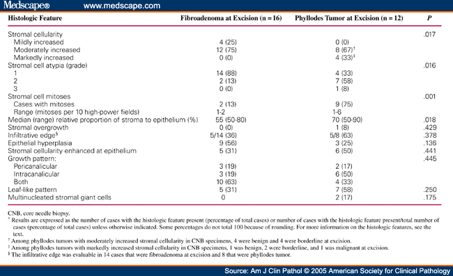
Fibroadenoma variants include cellular, complex, juvenile and myxoid forms. The cellular variant shows increased density of stromal cells within the architecture of a typical fibroadenoma, without significant stromal atypia, excess stromal mitotic activity or accentuated intracanalicularity (Fig. 4 ). The main differential diagnosis of the cellular fibroadenoma, especially on core biopsy, is the phyllodes tumour, which is distinguished by the presence of well-formed stromal fronds. In a long-term follow-up study conducted on a series of cellular fibroepithelial lesions that included 35 cellular fibroadenomas, none of which were widely excised, it was concluded that the recurrence rate of these tumours was low, without any phyllodes tumours diagnosed among the recurrences [ 11 ]. Genomically, cellular fibroadenomas possessed similar rates of mutations in the most commonly mutated genes MED12, KMT2D and RARA (49%, 13% and 13%) as conventional fibroadenomas (44%, 15% and 8%), indirectly supporting their classification with conventional fibroadenomas [ 12 ]. In contrast, the mutation spectrum of benign phyllodes tumours with which they resemble disclosed 62%, 14% and 17% abnormalities in the same set of genes, with a significant difference in the MED12 mutation rate. In addition, TERT promoter mutations were significantly higher in benign phyllodes tumours (32%) than in cellular (4%) and conventional (6%) fibroadenomas [ 12 ].
What is the difference between fibroadenoma and phyllodes?
The fibroadenoma is the commonest benign breast tumour in women, while the phyllodes tumour is rare and may be associated with recurrences, grade progression and even metastasis. The diagnosis of fibroadenoma is usually straightforward, with recognised histological variants such as the cellular, complex, juvenile and myxoid forms. The phyllodes tumour comprises benign, borderline and malignant varieties, graded using a constellation of histological parameters based on stromal characteristics of hypercellularity, atypia, mitoses, overgrowth and the nature of tumour borders. While phyllodes tumour grade correlates with clinical behaviour, interobserver variability in assessing multiple parameters that are potentially of different biological weightage leads to significant challenges in accurate grade determination and consequently therapy. Differential diagnostic considerations along the spectrum of fibroepithelial tumours can be problematic in routine practice. Recent discoveries of the molecular underpinnings of these tumours may have diagnostic, prognostic and therapeutic implications.
What is a fibroadenomas and phyllodes?
In summary, fibroadenomas and phyllodes are a fascinating group of fibroepithelial tumours that share not only morphological appearances but also genomic changes that underpin their pathogenesis.
What is a peri-epithelial condensation?
a Peri-epithelial or subepithelial stromal condensation is seen as an aggregation of stromal cells hugging the epithelium, which at low magnification may be discerned as a ‘shadow’ around the epithelial component. b Elongated clefts lined by benign epithelium may be a clue to more diagnostic phyllodal areas.
Is fibroepithelial lesions a neoplasm?
Breast fibroepithelial lesions are biphasic neoplasms composed of both epithelial and stromal components, comprising the common fibroadenoma and the less frequently occurring phyllodes tumour [ 1 ]. While the diagnosis of fibroadenoma is made relatively often, especially in core biopsies, the phyllodes tumour is a less commonly encountered ...
Which field of the fibroadenoma is stroma?
In the right field, stroma grows against, compresses and stretches the epithelium (intracanalicular pattern), while in the left field, stroma surrounds patent tubules reflecting the pericanalicular pattern. Full size image. A variety of histological changes can be seen in the fibroadenoma.
Is fibroadenomas on core biopsy?
Fibroadenomas are commonly diagnosed on core biopsy material. A question that is sometimes raised is whether a conclusion of fibroadenoma on core biopsy is accurate and reliable, and whether there should be concern for undersampling of a phyllodes tumour.
What is a neoplasm of the breast?
A type of connective tissue neoplasm typically arising from intralobular stroma of the breast. It is characterized by the rapid enlargement of an asymmetric firm mobile mass. Histologically, its leaf-like stromal clefts are lined by epithelial cells. Rare phyllodes tumor of the prostate is also known.
Can a fibroepithelial tumor recur after resection?
A benign, borderline, or malignant fibroepithelial neoplasm arising from the breast and rarely the prostate gland. It may recur following resection. The recurrence rates are higher for borderline and malignant phyllodes tumors. In borderline and malignant phyllodes tumors metastases to distant anatomic sites can occur.
Fibroepithelial tumors
Cite this page: Alexander M. Fibroadenomatoid change. PathologyOutlines.com website. https://www.pathologyoutlines.com/topic/breastfibroadenomatoidchange.html. Accessed January 3rd, 2022.
Fibroadenomatoid change
Cite this page: Alexander M. Fibroadenomatoid change. PathologyOutlines.com website. https://www.pathologyoutlines.com/topic/breastfibroadenomatoidchange.html. Accessed January 3rd, 2022.

Popular Posts:
- 1. icd-10 code for dilantin toxicity
- 2. icd 9 code for diabetes with microalbuminuria
- 3. icd-10 2015 code for acute st elevation (stemi) inferolateral myocardial infarction
- 4. 2019 icd 10 code for sclerotic lesion thoracic
- 5. icd-10-cm code for blood transfusion
- 6. icd 9 code for readmission algorithm
- 7. icd-10 code for cocaine use
- 8. icd 10 code for ocd behavior
- 9. icd 10 code for acute on chronic abdominal pain
- 10. icd 10 code for ashd