Short description: Macular cyst or hole. ICD-9-CM 362.54 is a billable medical code that can be used to indicate a diagnosis on a reimbursement claim, however, 362.54 should only be used for claims with a date of service on or before September 30, 2015.
What is the ICD 9 code for macular hole?
Macular Hole ICD-9 code: 362.54 A macular hole (MH) is a retinal break commonly involving the fovea. Idiopathic macular hole is the most common presentation. Risk factors include age, female gender, myopia, trauma, or ocular inflammation. Different findings can be observed depending the stage of the MH.
How do you diagnose a lamellar macular hole?
Differential diagnosis. OCT can be used to differentiate between true MH and conditions that may appear similar on clinical examination, including lamellar macular hole and pseudohole, among others. 2 Lamellar macular holes have an irregular contour with a break in the inner fovea without a break in the outer retina.
Is there a new classification for lamellar macular holes?
A new classification is needed to help clinicians in the management of lamellar macular holes. Some clinicians prefer to observe these clinical entities, especially when visual acuity is maintained or alterations of the photoreceptor layer are present.
What is the ICD 10 code for lamellar ichthyosis?
Lamellar ichthyosis. Q80.2 is a billable/specific ICD-10-CM code that can be used to indicate a diagnosis for reimbursement purposes.
What is the ICD-10 code for lamellar macular hole?
Macular cyst, hole, or pseudohole, unspecified eye H35. 349 is a billable/specific ICD-10-CM code that can be used to indicate a diagnosis for reimbursement purposes. The 2022 edition of ICD-10-CM H35. 349 became effective on October 1, 2021.
What is the ICD-10 code for macular hole left eye?
ICD-10 Code for Macular cyst, hole, or pseudohole, left eye- H35. 342- Codify by AAPC.
How do you describe a macular hole?
A macular hole is a rare eye condition that can blur the central vision you use to do everyday tasks like driving or reading. The macula is a small area in the center of the retina (the light-sensitive layer of tissue in the back of the eye).
What is a macular hole called?
A macular hole is a small break or hole in the central portion of the retina, called the macula. The macula is the central part of the retina which is responsible for distinguishing small details. Macular holes occur most frequently in healthy people and are most common in people in their 60s and 70s.
What is a lamellar hole?
Lamellar macular hole (LMH) is a vitreoretinal disorder characterized by an irregular foveal contour, a break in the inner fovea, dehiscence of the inner foveal retina from the outer retina, and the absence of a full-thickness foveal defect with intact foveal photoreceptors. The pathogenesis is only partially known.
What is full thickness macular hole?
Full thickness macular hole (FTMH) is a common maculopathy, which causes debilitating central vision loss and impairment of the quality of life of patients. It is usually idiopathic, but may be associated with trauma, high myopia and solar retinopathy.
Is a retinal hole the same as a macular hole?
A retinal hole is a small break or defect in the light-sensitive retina that lines the inside of the back of the eye. Retinal holes can occur anywhere in the retina. When a hole develops in the macula lutea (the most sensitive part of the central retina), it's called a macular hole.
Is a macular hole the same as macular degeneration?
A macular hole is not the same as macular degeneration, although they affect the same area of the eye and can sometimes both be present in the same eye. AMD is damage to the macula leading to the gradual loss of central vision.
Is a macular hole the same as a detached retina?
The size of the hole and its location on the retina determine how much it will affect a person's vision. A Stage III macular hole can destroy most central and detailed vision. If left untreated, a macular hole can lead to a detached retina. Detached retina is a serious condition that can result in severe vision loss.
How do you treat a lamellar hole?
Conclusion. In patients with lamellar macular hole, combination treatment with pars plana vitrectomy and internal limiting membrane peeling appears to be effective, but further studies are required to establish new treatment modalities for patients who do not have a satisfactory outcome from treatment.
What does a hole in the macula mean?
A macular hole within the eye's retina occurs when the nerve cells of the macula become separated from each other and pull away from the back surface of the eye, affecting vision. A macular hole can be successfully treated.
What is a stage 1 macular hole?
A stage 1A is a foveolar detachment characterized a loss of the foveal contour and a lipofuscin-colored spot. A stage 1B is a foveal detachment characterized by a lipofuscin-colored ring. Stage 2 MH is defined by a full thickness break < 400µm in size.
How do you heal a macular hole without surgery?
An untreated macular hole, left, and during treatment with the eye drops, right. Medicated drops may help close small macular holes over a two- to eight-week period, allowing some people to avoid surgery to fix the vision problem, a new study suggests.
What are the stages of macular hole?
There are three stages of a macular hole, foveal detachments (stage 1), partial-thickness holes (stage 2) and full-thickness holes (stage 3). Each stage can progress to the next if not treated. Surgery is over 95% effective for the treatment of macular holes.
How serious is a hole in your retina?
Retinal holes and tears are small breaks in the retina. The retina is light-sensitive tissue at the back of the eye. Usually holes and tears do not mean you will have serious vision problems right away. However, retinal holes and tears may cause problems if they allow fluid to seep behind the retina.
How long does it take to recover from macular hole surgery?
The total recovery time is several months. Patients will be asked to maintain face down positioning after surgery, from one to seven days, depending on a variety of patient-specific factors. Patients are on post-operative eye drops for a few weeks. The gas bubble gradually resorbs over two to eight weeks.
What are the stages of macular holes?
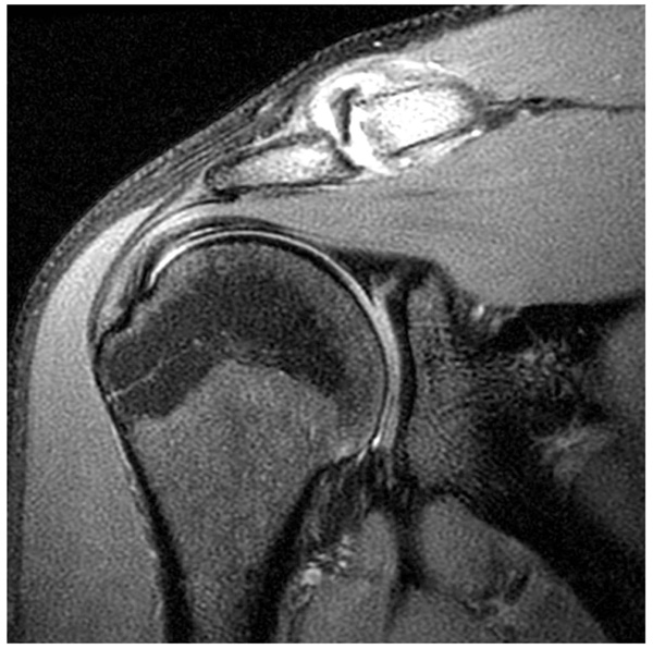
There are two main classification schemes for macular holes. Gass first described his clinical observations on the evolution of a macular hole: 1 Stage 1 MH, or impending MH, demonstrates a loss of the foveal depression. A stage 1A is a foveolar detachment characterized a loss of the foveal contour and a lipofuscin-colored spot. A stage 1B is a foveal detachment characterized by a lipofuscin-colored ring. 2 Stage 2 MH is defined by a full thickness break < 400µm in size. It might be eccentric with an inner layer “roof.” This can occur weeks to months following Stage 1 MHs. A further decline in visual acuity is also noted. In most cases, the posterior hyaloid has been confirmed to be still attached to the fovea on OCT analysis. 3 Stage 3 MH is further progression to a hole ≥400 µm in size. Nearly 100% of stage 2 MHs progress to Stage 3 and the vision further declines. A grayish macular rim often denotes a cuff of subretinal fluid. The posterior hyaloid is noted to be detached over the macula with or without an overlying operculum ( Figure 2 and 3 ). 4 Stage 4 MH is characterized by a stage 3 MH with a complete posterior vitreous detachment and Weiss ring.
Is Pars plana vitrectomy effective for MH?
There are no preventative measures for idiopathic MHs. Pars plana vitrectomy has not been clearly demonstrated to be effective in preventing MH formation.
Not Valid for Submission
362.54 is a legacy non-billable code used to specify a medical diagnosis of macular cyst, hole, or pseudohole. This code was replaced on September 30, 2015 by its ICD-10 equivalent.
Convert 362.54 to ICD-10
The following crosswalk between ICD-9 to ICD-10 is based based on the General Equivalence Mappings (GEMS) information:
Information for Medical Professionals
References found for the code 362.54 in the Index of Diseases and Injuries:
Information for Patients
The retina is a layer of tissue in the back of your eye that senses light and sends images to your brain. In the center of this nerve tissue is the macula. It provides the sharp, central vision needed for reading, driving and seeing fine detail.
ICD-9 Footnotes
General Equivalence Map Definitions The ICD-9 and ICD-10 GEMs are used to facilitate linking between the diagnosis codes in ICD-9-CM and the new ICD-10-CM code set. The GEMs are the raw material from which providers, health information vendors and payers can derive specific applied mappings to meet their needs.
What is a lamellar macular hole?
Lamellar macular holes are a vitreoretinal condition characterized by abnormalities in foveal contour with splitting of the neuroepithelium and often an intact photoreceptor layer. Recent developments in high-resolution imaging have increased our ability to study the details of the vitreoretinal int …
What are lamellar holes?
Lamellar macular holes are a vitreoretinal condition characterized by abnormalities in foveal contour with splitting of the neuroepithelium and often an intact photoreceptor layer. Recent developments in high-resolution imaging have increased our ability to study the details of the vitreoretinal interface and to distinguish between different forms of lamellar holes. A new classification is needed to help clinicians in the management of lamellar macular holes. Some clinicians prefer to observe these clinical entities, especially when visual acuity is maintained or alterations of the photoreceptor layer are present. Nevertheless, lamellar holes may sometimes progress, and visual acuity can deteriorate. On the other hand, surgical treatment may lead to positive anatomical and functional outcomes, but not without risks. This review provides a critical overview of the available data on lamellar macular holes, focusing on diagnosis and managing options.
What is the cause of macular holes?
Macular holes (MH) result in central vision loss, metamorphopsia, and a central scotoma. Idiopathic macular holes (IMHs) account for 83% of cases; they are found more commonly in females and are associated with increased age. Trauma is another cause, with traumatic macular holes (TMHs) comprising 5% to 8% of total cases. 1 It is important to consider TMH as a cause of vision loss following trauma, as it will inform medical and surgical management.
How to diagnose MH?
MH is diagnosed by means of a thorough history and clinical examination including slit-lamp biomicroscopy of the fundus with a dilated pupil. Symptoms usually include blurred central vision, inability to read or watch television, and distortion of the central vision with metamorphopsia. Visual acuity is decreased, and Amsler grid testing shows the central scotoma and metamorphopsia. At the slit lamp, a Watzke-Allen sign shows a break noted by the patient when a thin slit-lamp beam is focused across the MH. OCT provides the detailed imaging to make the diagnosis of full-thickness MH, which shows a break involving all the different layers of the retina.
What is the procedure to remove a MH?
Surgery. Surgical technique for MHs has evolved since the initial description by Kelly and Wendel in 1991. 7 Vitrectomy, membrane peeling, and the use of an intraocular tamponade, such as SF 6 gas, C 3 F 8 gas, or silicone oil, are utilized to relieve traction on the MH and to stimulate closure of the MH. Peeling of the internal limiting membrane (ILM) is performed by many vitreoretinal surgeons. This step often utilizes agents to assist in peeling including brilliant blue dye, indocyanine green dye, and triamcinolone.
Popular Posts:
- 1. icd 10 code for status post prostatectomy
- 2. icd 10 code for urethrorrhagia
- 3. icd 10 code for personal history of tubal ligation
- 4. icd 10 code for open wound right arm
- 5. icd 10 code for c1 hairline fracture
- 6. icd-10 code for benzodiazepine use disorder
- 7. icd-10 code for pain in left leg
- 8. icd 10 code for spider bite left leg
- 9. icd 10 cm code for 12 step group councling
- 10. icd-9 code for traumatic brain injury