What is the clinical margin of lentigo maligna?
First, the clinical margin of the lentigo maligna is outlined taking into account the clinical appearance with natural light, Wood’s lamp examination, and the histopathologic diagnosis of previous scouting biopsies.
Does lentigo maligna show up on R21?
With R21, nuclear expression of sAC is detected in almost 90% cases of lentigo maligna, but not in nevi. However, 25–30% of melanocytic hyperplasia in benign lentigines can show nuclear staining of sAC, which then needs to be distinguished from lentigo maligna with the hematoxylin and eosin stain [49].
How is lentigo maligna diagnosed?
Diagnosis The diagnosis of lentigo maligna is challenging, as the clinical presentation can be subtle and varied. Early detection of lentigo maligna relies on a high clinical suspicion index.
What stains are used in the diagnosis of lentigo maligna?
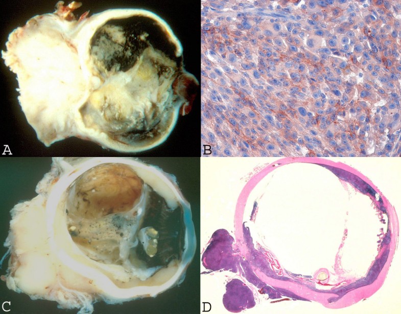
Immunohistochemical (IHC) stains are often used to aid in the diagnosis of lentigo maligna. Melanoma antigen recognized by T cells (MART-1 also known as Melan A) and microophthalmia transcription factor (MiTF) are the two stains regularly used at our institution. MiTF is expressed in the nucleus of melanocytes.

What is the diagnosis code for melanoma?
ICD-10-CM Code for Malignant melanoma of skin, unspecified C43. 9.
What is the ICD-10 code for melanoma face?
C43.39Malignant melanoma of other parts of face C43. 39 is a billable/specific ICD-10-CM code that can be used to indicate a diagnosis for reimbursement purposes. The 2022 edition of ICD-10-CM C43. 39 became effective on October 1, 2021.
What is the ICD 9 code for basal cell carcinoma?
ICD-9 code 173.31 for Basal cell carcinoma of skin of other and unspecified parts of face is a medical classification as listed by WHO under the range -MALIGNANT NEOPLASM OF BONE, CONNECTIVE TISSUE, SKIN, AND BREAST (170-176).
What is the code for Malignant melanoma of skin of scalp?
ICD-10-CM Code for Malignant melanoma of scalp and neck C43. 4.
What is lentigo maligna?
Lentigo maligna (LM) and lentigo maligna melanoma (LMM) are types of skin cancer. They begin when the melanocytes in the skin grow out of control and form tumors. Melanocytes are the cells responsible for making melanin, the pigment that determines the color of the skin.
What is the ICD-10 code for melanoma in situ?
D03. 9 is a billable/specific ICD-10-CM code that can be used to indicate a diagnosis for reimbursement purposes.
What is ICD-10 code for basal cell carcinoma?
ICD-10 code C44. 91 for Basal cell carcinoma of skin, unspecified is a medical classification as listed by WHO under the range - Malignant neoplasms .
How do you code basal cell carcinoma?
C44. 91 - Basal cell carcinoma of skin, unspecified | ICD-10-CM.
What is the CPT code for excision of basal cell carcinoma?
This type of excision would be most appropriately reported using the excision of malignant lesion including margins codes 11600- 11646.
How do you code metastatic melanoma?
VICC confirms that the correct code to assign for metastic melanoma at C4-C5 is C79. 5 Secondary malignant neoplasm of bone and bone marrow and that coding rules are not overridden to arrive at this code.
Which code represents excision Malignant lesion including margins eyelids 6.2 cm?
The excision of a malignant skin lesion including margins (procedure codes 11600-11646) will be considered medically necessary when a pathology report verifies the existence of a malignancy.
What is lentigo maligna?
Lentigo maligna is a melanocytic neoplasm occurring on sun-exposed skin, usually on the head and neck, of middle-aged and elderly patients. It is thought to represent the in situ phase of lentigo maligna melanoma. The ill-defined nature and potentially large size of lesions can pose significant diagnostic and treatment challenges. The goal of therapy is to cure the lesions in order to prevent development of invasive disease, and surgical excision is the treatment of choice to achieve clear histological margins. Nonsurgical treatment modalities have been reported; however, evidence is lacking to support their use. Age, general health, and comorbidities need to be taken into account when deciding the right treatment modality for each individual patient.
What is the treatment for lentigo maligna?
Radiation therapy . Radiotherapy, like imiquimod, is a noninvasive treatment option that has been used as a primary treatment for lentigo maligna in patients who are poor surgical candidates. Studies have used Grenz ray therapy for treatment of lentigo maligna [102–104].
What is the best biopsy for lentigo maligna?
Excisional biopsy is ideal for diagnosis of lentigo maligna [ 40 ]. In theory, excisional biopsy removes the whole clinical lesion down to subcutaneous fat with a 1–3 mm margin. This potentially allows for complete evaluation of depth and peripheral involvement. Excisional biopsy , however, is often not feasible for lentigo maligna because the lesions are typically ill-defined, widespread, and located in cosmetically sensitive areas. If the size of the lesion limits the ability to perform an excisional biopsy, scouting shave or punch biopsies can be performed ( Figure 1B ). Scouting biopsies should include samples from the darkest part, or most concerning part of the lesion, which will minimize the sampling error. They can also be taken from the periphery of the lesion to help delineate the peripheral margin involvement ( Figure 1B ).
Why is woods light used in lentigo maligna?
Since the true margins of lentigo maligna can exist far beyond the margins seen with visible light, the Wood’s light is used to improve margin delineation.
Where does lentigo maligna most commonly present?
Clinical presentation, risk factors, and genetics. Lentigo maligna most commonly presents on the head and neck region of elderly patients, with the highest incidence in the seventh and eighth decades of life.
Can you do an excisional biopsy on lentigo maligna?
Excisional biopsy, however, is often not feasible for lentigo maligna because the lesions are typically ill-defined, widespread, and located in cosmetically sensitive areas. If the size of the lesion limits the ability to perform an excisional biopsy, scouting shave or punch biopsies can be performed (Figure 1B).
Can lentigo maligna develop de novoor?
Lentigo maligna can develop de novoor within a pre-existing solar lentigo. Patients typically present with a chief complaint of a new, asymptomatic pigmented macule or patch on the head or neck region, or a freckle that has changed in size, shape, or color. Open in a separate window. Figure 1A.
What is the code for a primary malignant neoplasm?
A primary malignant neoplasm that overlaps two or more contiguous (next to each other) sites should be classified to the subcategory/code .8 ('overlapping lesion'), unless the combination is specifically indexed elsewhere.
What is secondary malignant melanoma?
Secondary malignant melanoma of skin. Superficial spreading malignant melanoma of skin. Clinical Information. A primary melanoma arising from atypical melanocytes in the skin.

Popular Posts:
- 1. icd-10-cm code for polycystic kidney disease, autosomal recessive
- 2. icd-10 code for wound infection unspecified
- 3. icd 10 code for granulation tissue
- 4. icd 10 code for renaldisease
- 5. icd 10 code for renal osteodystrophy
- 6. icd 10 code for high tsh
- 7. icd 10 code for non traumatic subarachnoid hemorrhage
- 8. icd 10 code for k44.9
- 9. icd 10 code for accidental overdose on seroquel
- 10. icd-9 code for hip replacement