Short description: Benign neoplasm skin NOS. ICD-9-CM 216.9 is a billable medical code that can be used to indicate a diagnosis on a reimbursement claim, however, 216.9 should only be used for claims with a date of service on or before September 30, 2015.
Benign neoplasm
A benign tumor is a mass of cells (tumor) that lacks the ability either to invade neighboring tissue or metastasize (spread throughout the body). When removed, benign tumors usually do not grow back, whereas malignant tumors are cancerous and sometimes do.
https://en.wikipedia.org › wiki › Benign_tumor
What is the ICD 10 code for neoplasm of unspecified breast?
2021 ICD-10-CM Diagnosis Code D24.9 Benign neoplasm of unspecified breast 2016 2017 2018 2019 2020 2021 Billable/Specific Code D24.9 is a billable/specific ICD-10-CM code that can be used to indicate a diagnosis for reimbursement purposes.
What is the ICD 10 code for excluded nevus?
When a type 2 excludes note appears under a code it is acceptable to use both the code (I78.1) and the excluded code together. blue nevus ( ICD-10-CM Diagnosis Code D22 flammeus nevus ( ICD-10-CM Diagnosis Code Q82.5 hairy nevus ( ICD-10-CM Diagnosis Code D22 melanocytic nevus ( ICD-10-CM Diagnosis Code D22
What is the ICD 9 code for syncope in breast NEC?
Short description: SYMPTOMS IN BREAST NEC. ICD-9-CM 611.79 is a billable medical code that can be used to indicate a diagnosis on a reimbursement claim, however, 611.79 should only be used for claims with a date of service on or before September 30, 2015.
What is the ICD 9 code for benign neoplasm of skin?
Benign neoplasm of skin, site unspecified. Short description: Benign neoplasm skin NOS. ICD-9-CM 216.9 is a billable medical code that can be used to indicate a diagnosis on a reimbursement claim, however, 216.9 should only be used for claims with a date of service on or before September 30, 2015.

What is the diagnosis code for nevus?
1.
What is the ICD-10 code for benign nevi?
I78.1I78. 1 is a billable/specific ICD-10-CM code that can be used to indicate a diagnosis for reimbursement purposes.
What is the ICD-10 code for atypical nevus?
D22. 9 is a billable/specific ICD-10-CM code that can be used to indicate a diagnosis for reimbursement purposes. The 2022 edition of ICD-10-CM D22.
What is diagnosis code D22 9?
Melanocytic nevi, unspecifiedICD-10 code D22. 9 for Melanocytic nevi, unspecified is a medical classification as listed by WHO under the range - Neoplasms .
What is an atypical nevus?
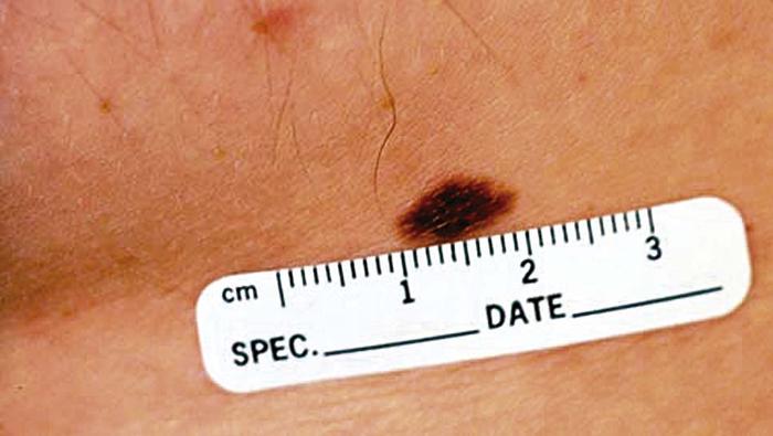
Atypical nevi, also known as dysplastic nevi, are benign acquired melanocytic neoplasms. Atypical nevi share some of the clinical features of melanoma, such as asymmetry, irregular borders, multiple colors, and diameter >5 mm (picture 1A). They occur sporadically or in a familial setting.
What is the ICD-10 code for pigmented nevi?
D22.9D22. 9 - Melanocytic nevi, unspecified | ICD-10-CM.
How do you code a dysplastic nevus?
ICD20 Dysplastic Nevi I would use D48. 5 for the dx of dysplastic nevi. Also, if the patient also has a hx of dysplastic nevi, don't forget to include Z86.
What is a non neoplastic nevus?
A abnormal, congenital formation or mark on the skin or neighboring mucosa that does not show neoplastic growth. [
What is a dysplastic nevus?
(dis-PLAS-tik NEE-vus) A specific type of nevus (mole) that looks different from a common mole. Dysplastic nevi are mostly flat and often larger than common moles and have borders that are irregular. A dysplastic nevus can contain different colors, which can range from pink to dark brown.
What is a compound nevus?
Listen to pronunciation. (KOM-pownd NEE-vus) A type of mole formed by groups of nevus cells found in the epidermis and dermis (the two main layers of tissue that make up the skin).
What is an acral nevus?
An acral nevus is a cutaneous condition of the palms, soles, fingers, or toes (peripheral body parts), characterized by a skin lesion that is usually macular or only slightly elevated, and may display a uniform brown or dark brown color, often with linear striations.
What is an intradermal nevus?
Intradermal melanocytic nevi are common, benign, pigmented skin tumors formed by proliferation of dermal melanocytes. A number of notable, uncommon changes may be observed in intradermal melanocytic nevi. In particular, their association with lymphatic invasion is an extremely rare phenomenon.
What is a nevus?
Nevus (or naevus, plural nevi or naevi, from nævus, Latin for "birthmark") is the medical term for sharply-circumscribed [1] and chronic lesions of the skin. These lesions are commonly named birthmarks and moles. Nevi are benign by definition.
Is nevus benign or malignant?
Nevi are benign by definition. Using the term nevus and nevi loosely, most physicians and dermatologists are actually referring to a variant of nevus called the "Melanocytic nevus", which are composed of melanocytes.
What is a mole on the skin?
A mole is a cluster of melanocytes and surrounding supportive tissue that usually appears as a tan, brown, or flesh-colored spot on the skin. The plural of nevus is nevi (nee-vye).
What is the plural of "nevus"?
The plural of nevus is nevi (nee-vye). A benign (not cancer) growth on the skin that is formed by a cluster of melanocytes (cells that make a substance called melanin, which gives color to skin and eyes). A mole is usually dark and may be raised from the skin.
What is a benign growth on the skin?
A benign growth on the skin (usually tan, brown, or flesh-colored) that contain s a cluster of melanocytes and surrounding supportive tissue. A neoplasm composed of melanocytes that usually appears as a dark spot on the skin. A nevus characterised by the presence of excessive pigment. A nevus containing melanin.
What is the code for a primary malignant neoplasm?
A primary malignant neoplasm that overlaps two or more contiguous (next to each other) sites should be classified to the subcategory/code .8 ('overlapping lesion'), unless the combination is specifically indexed elsewhere.
What is the table of neoplasms used for?
The Table of Neoplasms should be used to identify the correct topography code. In a few cases, such as for malignant melanoma and certain neuroendocrine tumors, the morphology (histologic type) is included in the category and codes. Primary malignant neoplasms overlapping site boundaries.
What chapter is functional activity?
Functional activity. All neoplasms are classified in this chapter, whether they are functionally active or not. An additional code from Chapter 4 may be used, to identify functional activity associated with any neoplasm. Morphology [Histology] Chapter 2 classifies neoplasms primarily by site (topography), with broad groupings for behavior, ...
What is the code for a primary malignant neoplasm?
A primary malignant neoplasm that overlaps two or more contiguous (next to each other) sites should be classified to the subcategory/code .8 ('overlapping lesion'), unless the combination is specifically indexed elsewhere.
What chapter is neoplasms classified in?
All neoplasms are classified in this chapter, whether they are functionally active or not. An additional code from Chapter 4 may be used, to identify functional activity associated with any neoplasm. Morphology [Histology] Chapter 2 classifies neoplasms primarily by site (topography), with broad groupings for behavior, malignant, in situ, benign, ...
What is the table of neoplasms used for?
The Table of Neoplasms should be used to identify the correct topography code. In a few cases, such as for malignant melanoma and certain neuroendocrine tumors, the morphology (histologic type) is included in the category and codes. Primary malignant neoplasms overlapping site boundaries.
Can multiple neoplasms be coded?
For multiple neoplasms of the same site that are not contiguous, such as tumors in different quadrants of the same breast, codes for each site should be assigned. Malignant neoplasm of ectopic tissue. Malignant neoplasms of ectopic tissue are to be coded to the site mentioned, e.g., ectopic pancreatic malignant neoplasms are coded to pancreas, ...
Popular Posts:
- 1. icd 10 code for hyper daytime somnolence
- 2. billable icd 9 code for history of gout
- 3. icd 10 code for thigh abscess
- 4. icd code for anaphylaxis
- 5. icd code for neck strain
- 6. icd 10 code for thoracic diskitis
- 7. icd 10 code for status post bladder surgery
- 8. icd 10 code for right hand splint
- 9. icd 10 code for genital fungal infection
- 10. icd 10 cm code for cardiomegaly